. 2016 Mar;24(2):328-36.
doi: 10.1111/wrr.12411. Epub 2016 Mar 10.

PKM2 released by neutrophils at wound site facilitates early wound healing by promoting angiogenesis

Affiliations

PKM2 released by neutrophils at wound site facilitates early wound healing by promoting angiogenesis

Yinwei Zhang et al. Wound Repair Regen. 2016 Mar.

Abstract

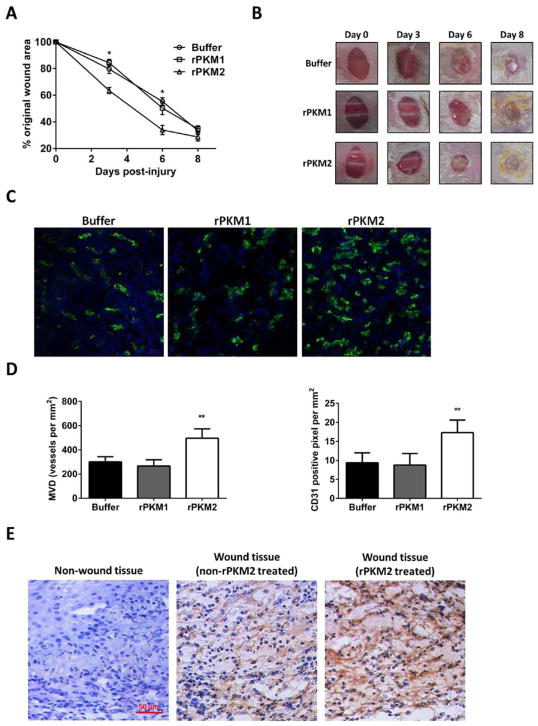
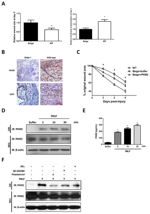
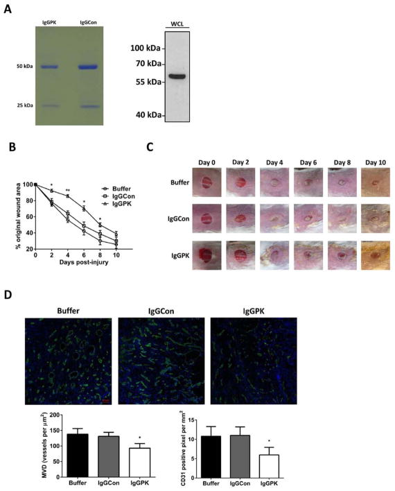
Neutrophils infiltration/activation following wound induction marks the early inflammatory response in wound repair. However, the role of the infiltrated/activated neutrophils in tissue regeneration/proliferation during wound repair is not well understood. Here, we report that infiltrated/activated neutrophils at wound site release pyruvate kinase M2 (PKM2) by its secretive mechanisms during early stages of wound repair. The released extracellular PKM2 facilitates early wound healing by promoting angiogenesis at wound site. Our studies reveal a new and important molecular linker between the early inflammatory response and proliferation phase in tissue repair process.


Conflict of interest statement

Conflict of Interest: All authors declare no conflict of interests related to publish of the article.

Figures

Figure 1
Figure 1
Extracellular PKM2 promotes wound healing. (A) The wound closure of CD-1 mice that were treated by the indicated agents. The wound closure is presented as % of original wound area, [(Original wound area - wound area)/original wound area]. (B) Representative pictures of wound areas at different days of treatments by indicated agents. (C) Representative images of immunostaining of wound tissue sections with antibodies against mouse CD31 (Green). The blues are DAPI stains. (D) Quantitative analyses of CD31 staining of the wound sections by manually counting using the software imaging-J. MVD (Left) is vessel density per mm2. The CD31 positive pixel (Right) is measured per mm2. The quantification was average of randomly selected 4 fields in randomly selected 3 sections. (E) Representative images of IHC staining of tissue sections from wound or un-wound area with/without application of rPKM2 using anti-PKM2 antibody. Blue is hematoxylin staining. Error bars in (A) and (D) are standard deviations from measurements of six mice.
Figure 2
Figure 2
Activated neutrophils release PKM2 at the wound site. (A, B) Representative images of IHC staining of tissue sections from wound or un-wound areas using antibodies against PKM2 or Ly6G, a neutrophil marker. (A) Sections were prepared from tissues of 3rd day post wound induction and stained by anti-PKM2 antibody. On the right is call-out enlargement of the indicated area. (B) Sections were prepared from wound tissues of different days (indicated) after wound induction. (C) Quantitation of extracellular PKM2 vs Ly6G stains in (B). Quantitations were done by using Frida software. Randomly selected three fields were analyzed. For quantitation of extracellular PKM2, extracellular PKM2 stains were manually selected in each view field and quantitated by the software. Error bars are standard deviations from measurements of five sections from five mice. (D) Representative images of IHC staining of tissue sections from wound areas on day 0 and day 3 using antibody against F4/80, a macrophage marker. Blue in (A), (B), and (D) is hematoxylin staining.
Figure 3
Figure 3
PKM2 present in secretive vesicles of activated neutrophils. (A, B) Levels of PKM2 in culture medium (CM) of neutrophils treated with indicated agents were analyzed by immunoblot (A) of PKM2 (IB:PKM2) or ELISA analyses (B) using goat anti-human PKM2 as capture antibody and rabbit monoclonal anti-PKM2 as detection antibody. Immunoblot of PKM2 (IB:PKM2) and β-actin (IB:β-actin) in cell lysate (WCL) in (A) were loading controls indicate amounts of neutrophils used in each experiment. Damn is Damnacathal. Error bars in (B) are standard deviations from five independent measurements. (C) Presence of PKM2 in each fraction of sucrose gradient of extracts of activated (bottom) and inactivated (upper) neutrophils (10 ml in total) was analyzed by immunoblot of PKM2 (IB:PKM2). Numbers on top indicate fraction number (1 ml each fraction from top down). Immunoblot of cd11b (IB:cd11b) indicate the fractions containing neutrophil granules (fractions 13–19). Immunoblot of β-actin (IB:β-actin) indicate general cytosolic fractions (fractions 1–10).
Figure 4
Figure 4
Activated neutrophils release PKM2 by secretive mechanism. (A) The wound closure (left) and granulation tissue thickness (right) of beige-J (Beige) mice or control mice (WT) at day three after wound induction. The wound closure granulation tissue thickness is presented as relative wound areas and relative thickness of granulation tissue respectively by define the average of wound area and granulation tissue thickness of Beige-J mice as 1. Error bars are standard deviations from measurements of six mice. (B) Representative images of IHC staining of sections from wound areas of beige-J (Beige-J) mice or control mice of the same strain without gene mutations (wild-type) using antibodies against PKM2 or Ly6G. Sections were prepared from tissues of the 3rd days post wound induction. Blue staining is hematoxylin staining. (C) The wound closure of beige-J (Beige) mice or control mice (WT) that were treated by rPKM2 or buffer. The wound closure is presented as % of original wound area as in Figure 1A. (D) and (E) Levels of PKM2 in culture medium (CM) of neutrophils treated with fMLP for different time (indicated) were analyzed by immunoblot (D) of PKM2 (IB:PKM2) or ELISA analyses (E) using goat anti-human PKM2 as capture antibody and rabbit monoclonal anti-PKM2 as detection antibody. (F) Levels of PKM2 in culture medium (CM) of neutrophils treated with indicated agents were analyzed by immunoblot of PKM2 (IB:PKM2). Immunoblot of PKM2 (IB:PKM2) and β-actin (IB:β-actin) in cell lysate (WCL) in (D, F) were loading controls indicate amounts of neutrophils used in each experiment. Error bars in (E) are standard deviations of five independent experiments, in (A, C) are standard deviations of experiment from five mice.
Figure 5
Figure 5
Effects of PKM2 antibody on wound closure and angiogenesis at wound site. (A) (Left) IgG purified from monoclonal antibody IgGPK or rabbit pre-immuno serum (IgGCon) by protein G/A beads. (Right) Specific recognition of PKM2 by the purified IgG of IgGPK in SW480 cell extracts. (B) Wound closure of CD-1 mice that were treated by topical application of indicated agents was monitored. The wound closure is presented as the % of original wound area. Error bars are standard deviations from measurements of six mice. (C) Representative pictures of wound areas at different days of treatment by indicated agents. (D) (Upper) Representative images of immunostaining of wound tissue sections with antibodies against mouse CD31 (Green). The blues are DAPI stains. The wounds were treated by the indicated agents. (Bottom) Quantitative analyses of CD31 staining of the wound sections by manually counting using the software imaging-J. MVD (bottom, left) is vessel density per mm2. The CD31 positive pixel (bottom, right) is measured per mm2. The quantification was average of randomly selected 4 fields in randomly selected 3 sections. Error bars are standard deviations from measurements of six mice.

References

    1. Singer AJ, Clark RA. Cutaneous wound healing. N Engl J Med. 1999;341(10):738–46. - PubMed
    1. Diegelmann RF, Evans MC. Wound healing: an overview of acute, fibrotic and delayed healing. Front Biosci. 2004;9:283–9. - PubMed
    1. Broughton G, 2nd, Janis JE, Attinger CE. Wound healing: an overview. Plast Reconstr Surg. 2006;117(7 Suppl):1e-S–32e-S. - PubMed
    1. Gillitzer R, Goebeler M. Chemokines in cutaneous wound healing. J Leukoc Biol. 2001;69(4):513–21. - PubMed
    1. Artlett CM. Inflammasomes in wound healing and fibrosis. J Pathol. 2013;229(2):157–67. - PubMed

Substances