. 2016 May;59(1):1-17.
doi: 10.1007/s12031-015-0711-6. Epub 2016 Jan 25.

Neuronal Hemoglobin Expression and Its Relevance to Multiple Sclerosis Neuropathology

Affiliations

Neuronal Hemoglobin Expression and Its Relevance to Multiple Sclerosis Neuropathology

Nolan Brown et al. J Mol Neurosci. 2016 May.

Abstract

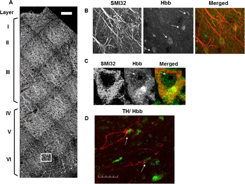
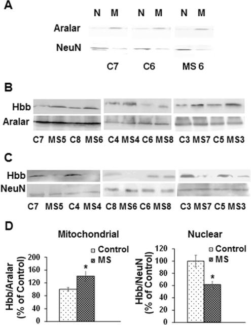
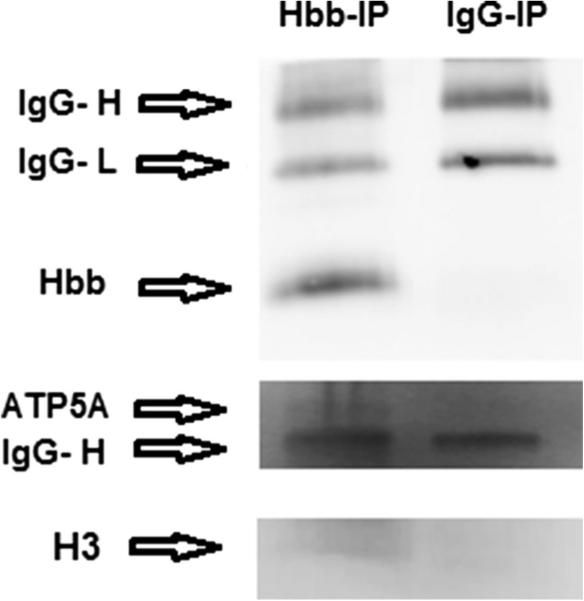
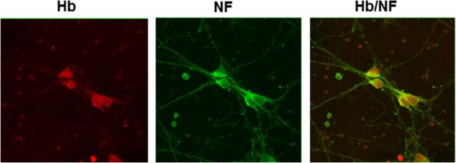
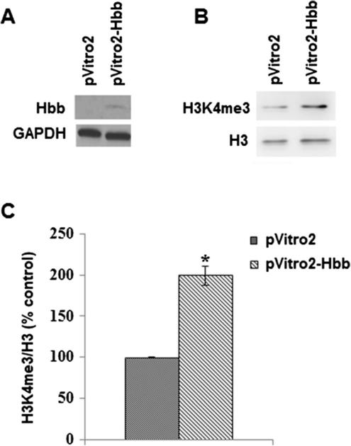
Multiple sclerosis (MS) is characterized by demyelination and progressive neurological disability. Previous studies have reported defects to mitochondria in MS including decreased expression of nuclear encoded electron transport chain subunit genes and inhibition of respiratory complexes. We previously reported increased levels of the hemoglobin β subunit (Hbb) in mitochondrial fractions isolated from postmortem MS cortex compared to controls. In the present study, we performed immunohistochemistry to determine the distribution of Hbb in postmortem MS cortex and identified proteins which interact with Hbb by liquid chromatography tandem mass spectrometry (LC-MS/MS). We found that Hbb was enriched in pyramidal neurons in internal layers of the cortex and interacts with subunits of ATP synthase, histones, and a histone lysine demethylase. We also found that Hbb is present in the nucleus and that expression of Hbb in SH-SY5Y neuroblastoma cells increased trimethylation of histone H3 on lysine 4 (H3K4me3), a histone mark that regulates cellular metabolism. These data suggest that Hbb may be a part of a mechanism linking neuronal energetics with epigenetic changes to histones in the nucleus and may provide neuroprotection in MS by supporting neuronal metabolism.

Keywords: Hemoglobin expression; Histone methylation; Mass spectrometry; Mitochondrial genes; Multiple sclerosis; Pyramidal neurons.


Figures

Fig. 1
Fig. 1
Cortical distribution of Hbb expressing pyramidal neurons in MS cortex. a Representative confocal image from MS motor cortex immunostained with antibodies to neurofilament (SMI32) and Hbb. Sequential ×20 confocal images were acquired from the pial surface extending through cortical layer VI and stitched into a single image. Cortical layers were visualized by SMI32 staining shown in grayscale. Scale bar represents 100 μm. b Panel shows the boxed region in A at higher magnification. SMI32 and Hbb immunoreactivity can be seen in pyramidal neurons denoted by arrows. SMI32 and Hbb channels are shown in grayscale and the merged image on the right shows colocalization of SMI32 (red fluorescence) and Hbb (green fluorescence). SMI32 and Hbb positive cells were counted in cortical tissue sections from five MS brains. We found that on average 33 % of SMI32+ cells were also immunoreactive for Hbb. In external layers of the cortex (layers I–III), 22 % of SMI32+ neurons were Hbb+. In cortical layers IV–VI, the internal cortical layers, 42 % of SMI32 neurons also expressed Hbb. c Hbb expression is observed in the nucleus but not in the nucleolus (denoted by arrows) in pyramidal cells. d Representative confocal image showing an MS cortical section immunostained with antibodies to TH (red fluorescence) and Hbb (green fluorescence). Arrows denote TH positive axons appearing to contact cell bodies of Hbb positive cells. Scale bar represents 30 μm
Fig. 2
Fig. 2
The subcellular localization of Hbb is altered in MS samples. a Representative Western blot demonstrating the purity of mitochondrial and nuclear fractions isolated from control and MS postmortem cortical gray matter. Nuclear (N) and mitochondrial (M) fractions isolated from control and MS samples were blotted with antibodies to the neuronal nuclear marker NeuN and the mitochondrial marker aralar. b Representative Western blots for Hbb and aralar in mitochondrial fractions isolated from MS cortical samples compared to controls. c Representative Western blots for Hbb and NeuN show decreased Hbb in MS nuclear fractions compared to controls. d Quantitation shows that average Hbb levels are increased in mitochondrial fractions and decreased in nuclear fractions isolated from MS cortical gray matter compared to controls. Error bars represent SEM. *p < 0.05
Fig. 3
Fig. 3
Confirmation of proteins interacting with Hbb that were identified by mass spectrometry. Western blotting shows that Hbb, ATP5A1, and histone H3 were pulled down in the IP experiment with the Hbb antibody but not with the non-specific IgG antibody. Arrows denote bands for Hbb (16 kDa), ATP5A1 (55 kDa), and histone H3 (13 kDa) on separate blots. Because this protein was immunoprecipitated with an Hbb antibody, IgG heavy and light chains (IgG-H and IgG-L) are present at 50 and ~25 kDa
Fig. 4
Fig. 4
Hemoglobin is expressed in cultured rat primary neurons. Cultured neuronal cells were immunostained with antibodies to hemoglobin (Hb) and neurofilament (NF). The merged image shows Hb (red fluorescence) and NF (green fluorescence) colocalization
Fig. 5
Fig. 5
Hbb expression regulates histone H3 trimethylation. a Western blotting shows that the Hbb subunit is expressed in SH-SY5Y cells transfected with pVitro2-Hbb. b Representative Western blot demonstrates that levels of H3K4me3 are increased in nuclear extracts isolated from SH-SY5Y cells transfected with pVitro2-Hbb compared to control cells transfected with vector alone. c Quantitation for H3K4me3 levels in SH-SY5Y cells transfected with either empty vector pVitro2 or pVitro2-Hbb was performed by densitometry from two separate experiments. Error bars represent SEM, *p < 0.05

References

    1. Bellelli A, Brunori M, Miele AE, Panetta G, Vallone B. The allosteric properties of hemoglobin: insights from natural and site directed mutants. Curr Protein Pept Sci. 2006;7:17–45. - PubMed
    1. Benavides-Piccione R, DeFelipe J. Distribution of neurons expressing tyrosine hydroxylase in the human cerebral cortex. J Anat. 2007;211(2):212–22. - PMC - PubMed
    1. Biagioli M, Pinto M, Cesselli D, Zaninello M, Lazarevic D, Roncaglia P, Simone R, Vlachouli C, Plessy C, Bertin N, Beltrami A, Kobayashi K, Gallo V, Santoro C, Ferrer I, Rivella S, Beltrami CA, Carninci P, Raviola E, Gustincich S. Unexpected expression of α- and β-globin in mesencephalic dopaminergic neurons and glial cells. Proc Natl Acad Sci U S A. 2009;106:15454–15459. - PMC - PubMed
    1. Bjartmar C, Kidd G, Mörk S, Rudick R, Trapp BD. Neurological disability correlates with spinal cord axonal loss and reduced N-acetyl-aspartate in chronic multiple sclerosis patients. Ann Neurol. 2000;48:893–901. - PubMed
    1. Bö L, Geurts JJ, Mörk SJ, van der Valk P. Grey matter pathology in multiple sclerosis. Acta Neurol Scand Suppl. 2006;183:48–50. - PubMed

MeSH terms

LinkOut - more resources