. 2016 Jan 26:11:11.
doi: 10.1186/s13024-016-0074-7.

A Fluorescent Oligothiophene-Bis-Triazine ligand interacts with PrP fibrils and detects SDS-resistant oligomers in human prion diseases

Affiliations

A Fluorescent Oligothiophene-Bis-Triazine ligand interacts with PrP fibrils and detects SDS-resistant oligomers in human prion diseases

Thibaut Imberdis et al. Mol Neurodegener. .

Abstract

Background: Prion diseases are characterized by the accumulation in the central nervous system of an abnormally folded isoform of the prion protein, named PrP(Sc). Aggregation of PrP(Sc) into oligomers and fibrils is critically involved in the pathogenesis of prion diseases. Oligomers are supposed to be the key neurotoxic agents in prion disease, so modulation of prion aggregation pathways with small molecules can be a valuable strategy for studying prion pathogenicity and for developing new diagnostic and therapeutic approaches. We previously identified thienyl pyrimidine compounds that induce SDS-resistant PrP(Sc) (rSDS-PrP(Sc)) oligomers in prion-infected samples.

Results: Due to the low effective doses of the thienyl pyrimidine hits, we synthesized a quaterthiophene-bis-triazine compound, called MR100 to better evaluate their diagnostic and therapeutic potentials. This molecule exhibits a powerful activity inducing rSDS-PrP(Sc) oligomers at nanomolar concentrations in prion-infected cells. Fluorescence interaction studies of MR100 with mouse PrP fibrils showed substantial modification of the spectrum, and the interaction was confirmed in vitro by production of rSDS-oligomer species upon incubation of MR100 with fibrils in SDS-PAGE gel. We further explored whether MR100 compound has a potential to be used in the diagnosis of prion diseases. Our results showed that: (i) MR100 can detect rSDS-oligomers in prion-infected brain homogenates of various species, including human samples from CJD patients; (ii) A protocol, called "Rapid Centrifugation Assay" (RCA), was developed based on MR100 property of inducing rSDS-PrP(Sc) oligomers only in prion-infected samples, and avoiding the protease digestion step. RCA allows the detection of both PK-sensitive and PK-resistant PrP(Sc) species in rodents samples but also from patients with different CJD forms (sporadic and new variant); (iii) A correlation could be established between the amount of rSDS-PrP(Sc) oligomers revealed by MR100 and the duration of the symptomatic phase of the disease in CJD patients; and (iv) Bioassay experiments showed that MR100 can trap prion infectivity more efficiently than P30 drug.

Conclusions: MR100 is a powerful tool not only for studying the prion aggregation pathways regarding oligomeric and sPrP(Sc) species, but also for developing alternative methods for the detection of prion-infected samples. Considering our bioassay results, MR100 is a promising molecule for the development of prion decontamination approaches.


Figures
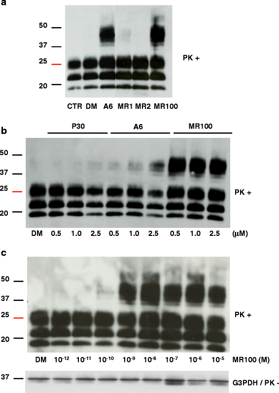
Fig. 1
Fig. 1
MR100 has a stronger PrPSc oligomer-inducing activity in prion-infected N2a58/22 L cells than P30 and A6. a Effect of the newly synthesized MR1, MR2 and MR100 compounds in prion-infected N2a58/22 L cells. Cells were left untreated (CTR) or incubated with 20 μM of A6 (positive control), MR1 and MR2 (synthesis intermediates), MR100, or 20 μL DMSO (DM) for 4 days. Protein lysates were analyzed by immunoblotting with the SAF mix (a mixture of the anti-PrP SAF60, SAF69 and SAF70 monoclonal antibodies) after proteinase K (PK) digestion. b Comparison of the oligomer-inducing activity of P30, A6 and MR100. Prion-infected N2a58/22 L cells were incubated with 0.5, 1 or 2.5 μM of each compound, or 20 μL DMSO (DM) for 4 days. Protein lysates were then analyzed by immunoblotting with the SAF mix after PK digestion. c MR100 dose-response curve in prion-infected N2a58/22 L cells. Successive dilutions of MR100 in DMSO were used to obtain final concentrations ranging from 10-12M (1pM) to 10-5M (10 μM). Cells were incubated for 4 days and at confluence they were lysed. Protein lysates were analyzed by immunoblotting with the SAF mix after PK digestion according to the previously described protocol [16, 30]. Loading control was performed with antibodies against glyceraldehyde-3-P dehydrogenase (G3PDH) and before proteinase K digestion. Molecular masses (20–50 kDa) are indicated on the left side of the panels
Fig. 2
Fig. 2
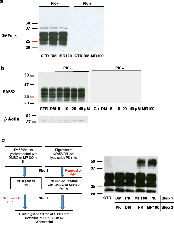
MR100 did not induce SDS resistant PrPC oligomers. a Parental non-infected cells, N2a58, were left untreated (CTR) or incubated with 20 μM of MR100 or 20 μL DMSO (DM) for 4 days. Protein lysates were analyzed by immunoblotting with the SAF mix before or after PK digestion. b N2a58 cell lines were left untreated (CTR) or incubated with various concentration of MR100 from 5 to 40 μM or 40 μL of DMSO (DM) for 4 days. Protein lysates were analyzed by immunoblotting with the SAF 32 before or after PK digestion. Loading control was performed with antibodies against β actin and before proteinase K digestion. c Schematic representation of the protocols used to test if PrPC isoforms are part of rSDS-oligomers (Left panel). First step, N2a58/22 L lysates were incubated with 20 μM of MR100 or with proteinase K to eliminate PrPC, then in the second step, MR100-exposed lysates were digested with proteinase K, while proteinase K digested samples were incubated with MR100. PrPSc species were then analyzed by western blotting. Western blot analysis of the samples processed according to the two different protocols using the SAFmix of anti-PrP antibodies (Right panel). CTR, untreated samples, digested by proteinase K; DM, DMSO. Molecular masses (20–50 kDa) are indicated on the left side of the panels
Fig. 3
Fig. 3
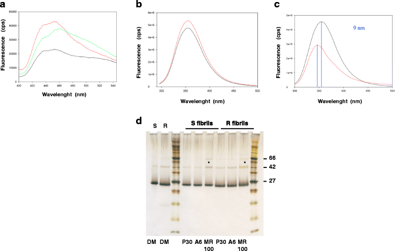
Fluorescence interaction studies between MR100 and PrP. a Fluorescence studies of MR100 compound incubated with purified recombinant MoPrP(23-230) proteins. 4 μM of MoPrP(23-230), either soluble or fibrillar, were incubated with 50 μM of MR100 in 1 % DMSO, 50 mM MES buffer pH6, during 2 h at 25 °C. Emission spectra between 400 and 550 nm were recorded by exciting at 470 nm: 50 μM of MR100 (black), 50 μM of MR100 + α-soluble MoPrP(23-230) (red), 50 μM of MR100 + fibrils of MoPrP(23-230) (green). b-c Fluorescence of tryptophan and tyrosine residues of soluble MoPrP(23-230) alone (black), or incubated with 1 % DMSO, 50 mM MES buffer pH6, during 2 h at 37 °C (red) (b). Fluorescence of tryptophan and tyrosine residues of soluble MoPrP(23-230) alone (black), or incubated with 50 μM of MR100 in 1 % DMSO, 50 mM MES buffer pH 6, during 2 h at 37 °C (red) (c). All spectra were recorded at 290 nm. d Hamster-S or -R fibrils at a concentration of 4 μM were incubated with solvent alone (1 % DMSO, 50 mM MES buffer pH6), or 40 μM of P30, A6, or MR100 compounds in 1 % DMSO, 50 mM MES buffer pH6, during 2 h at 25 °C. Samples were then mixed with loading buffer, boiled for 15 min at 90 °C and loaded on a 12 % SDS-PAGE gel. Proteins in the gel were revealed by silver staining. Molecular weight markers indicated: 27, 42 and 66 kDa. Asterisks showed dimer and trimer bands that are increased following incubation with MR100 compound
Fig. 4
Fig. 4
MR100 shows oligomer-inducing activity in brain homogenates from prion-infected rodents. a MR100 oligomer-inducing activity was tested using freshly homogenized rodent brain tissues infected with the 22 L (mice) or the 263 K prion strain (hamsters). Fifty μL of 10 % mouse or hamster brain homogenates were diluted in 300 μL PBS/2 % Sarkosyl, incubated with 1.5 mM MR100 (corresponding to 150 μL of 5 mM MR100) at room temperature for 1 h or with 150 μL of DMSO as control, and then digested with 20 μg/ml PK at a ratio of 1:50 (PK/proteins). PK digestion was stopped by addition of a cocktail of protease inhibitors (Complete), before analysis of rPrPSc by western blotting with the SAF mix according to the previously described protocol [16]. CTR: untreated 22 L- or 263 K-infected brain homogenates (negative control); DM: 22 L- or 263 K-infected brain homogenates incubated with DMSO. b Comparison of P30 and MR100 oligomer-inducing activity on the 22 L prion strain, before and after proteinase K digestion. Fifty μL of 10 % 22 L-infected brain homogenates were diluted in 350 μL PBS/2 % Sarkosyl, incubated with 1 mM MR100 or P30 (corresponding to 100 μL of 5 mM MR100 or P30), at room temperature for 1 h. Then, aliquots of 30 μL were taken before addition of proteinase K, to perform western blot (PK-) probed with SAF mix, but also with anti-β-actin antibodies as loading controls. The rest of the sample was then digested with 20 μg/ml PK at a ratio of 1:50 (PK/proteins) (PK+). The reaction was stopped by addition of the protease inhibitor cocktail, before analysis of rPrPSc by western blotting with the SAF mix as in A. DM: 22 L-infected brain homogenates incubated with 100 μL DMSO. Molecular masses (20–50 kDa) are indicated on the left side of the panels
Fig. 5
Fig. 5
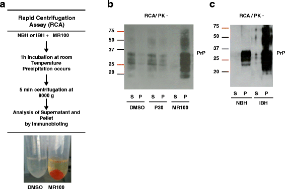
A MR100-based assay can differentiate between prion-infected and normal brain homogenates without proteinase K digestion. a Schematic description of the RCA protocol to test brain homogenates without PK digestion. Brain tissues were freshly homogenized in microbead-containing tubes. Normal brain homogenates (NBH) or prion-infected brain homogenates (IBH) were incubated with MR100 for 1 h, at room temperature, leading to a precipitation of PrP isoforms. After a short centrifugation step, the pellet with MR100 (orange tube) concentrates PrP isoforms, whereas no pellet is detectable with DMSO. b Comparison of DMSO, P30 and MR100 precipitation capabilities using the RCA protocol. Fifty μL of 10 % 22 L infected brain homogenates were diluted in 300 μL of PBS/2 % sarcosyl and incubated using either 1.5 mM of P30 or MR100 or an equivalent volume of the solvent alone (DMSO), at room temperature for 1 h. Then, samples were centrifuged at 8000 g for 5 min. Supernatants (S) were collected and 30 μL of each supernatant was mixed with an equivalent volume of 2X loading buffer. Pellets (P) were resuspended in 30 μL PBS/2 % Sarcosyl, and mixed with an equal volume of 2X loading buffer. Thirty microliters of each sample were loaded on 12 % Bis-Tris gels (Criterion, Biorad) and immunoblotting was carried out with the SAF mix according to standard procedures [16]. The samples (S/P) were analyzed by western blotting using SAF mix anti-PrP antibodies. c Comparison of infected versus non-infected brain homogenates processed with the RCA protocol. Fifty microliters of 10 % freshly homogenized brain tissues from normal (NBH) or 22 L prion-infected (IBH) mice were processed according to the RCA protocol described in A and B. Thirty microliters of supernatant (S) or pellet (P) were loaded on 12 % Bis-Tris gels (Criterion, Biorad) and immunoblotting was carried out with the SAF mix as described above. Molecular masses (20–75 kDa) are indicated on the left side of the panels
Fig. 6
Fig. 6
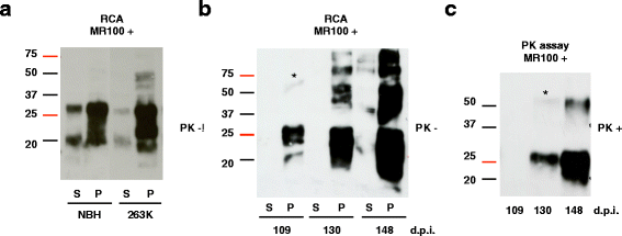
RCA can detect rSDS-PrPSc oligomers at early stages of the disease. a Fifty microliters of 10 % hamster brain homogenates (NBH or infected with the 263 K prion strain) were diluted in 300 μL of PBS/2 % Sarkosyl, incubated with 1.5 mM MR100 at room temperature for 1 h and then centrifuged at 8000 g for 5 min. Supernatants (S) were collected and 30 μL of each supernatant were mixed with an equivalent volume of 2X loading buffer. Pellets (P) were resuspended in 30 μL PBS/2 % Sarcosyl, and mixed with 30 μL of 2X loading buffer. Thirty microliters of each sample were loaded on 12 % Bis-tris gels (Criterion, Biorad) and western blotting was carried out with the SAF mix according to standard procedures [16]. Molecular masses (20–75 kDa) are indicated on the left side of the panels. b Hamster brain tissues were collected at various days post-infection (d.p.i.), as indicated, and freshly homogenized tissues were processed according to the RCA protocol using MR100 and analyzed by immunoblotting as described above. c To compare the RCA and the PK test, the same hamster brain homogenates (at 109, 130 and 148 d.p.i.) were incubated with MR100 at room temperature for 1 h, then digested with 20 μg/mL of proteinase K and processed as described in the legend to Fig. 4a. The asterisk in b and c indicates the position of oligomer traces
Fig. 7
Fig. 7
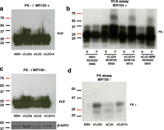
rSDS-PrPSc oligomers are detected in patients with new variant CJD (vCJD) when tested with RCA. a-b Frozen, homogenized brain samples from two patients with vCJD, one patient with sCJD (codon 129 M/M genotype) (positive control) and one healthy control (NBH) were from NIBSC. Each sample was identified by the number attributed by the NIBSC. The RCA assay was carried out as before (see legend to Fig. 5) but adapted to human samples: 50 μL of 10 % brain homogenates in 100 μL PBS/2 % Sarkosyl were incubated with 2 mM MR100 for 2 h. Before centrifugation, 30 μL was collected and mixed with 30 μL of 2X loading buffer for immunoblotting analysis (a). The rest of the sample was centrifuged at 11,000 g for 5 min, and supernatants (S) and pellets (P) were immunoblotted with the SAFmix (b) [16]. c-d For comparison, the same brain homogenates (50 μL of 10 % brain homogenates in 100 μL PBS/2 % Sarkosyl) were processed with the classical proteinase K digestion assay without MR100. Samples were analyzed before (c), and after proteinase K digestion (125 μg/mL PK at 37 °C for 1 h) (d). The reaction was stopped by addition of a protease inhibitor cocktail, before analysis of rPrPSc by western blotting with the SAF mix. Molecular masses (20–75 kDa) are on the left side of the panels
Fig. 8
Fig. 8
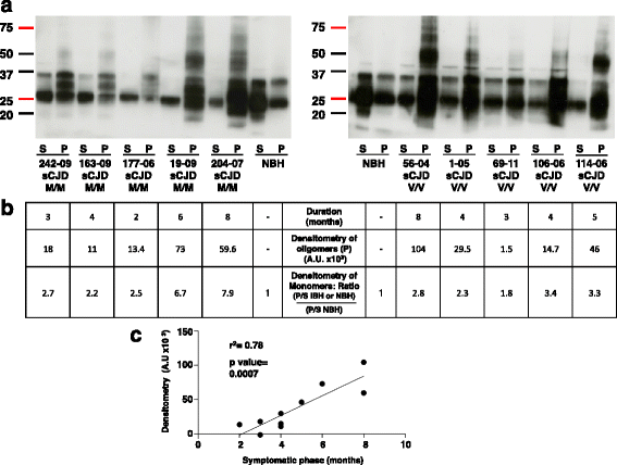
RCA efficiently detects rSDS-PrPSc oligomers in patients with sporadic Creutzfeldt-Jakob disease (sCJD). a Freshly homogenized human brain tissues from patients with sCJD (codon 129 Met/Met or Val/Val polymorphisms) were analyzed using RCA. The number on the figures corresponds to the identification numbers attributed by the tissue bank. The brain homogenate from a healthy control (NBH, the same as in Fig. 7) was used as negative control. 50 μL of 10 % human brain homogenates in 100 μL PBS/2 % Sarkosyl were incubated with 2 mM MR100 at room temperature for 2 h and centrifuged at 11,000 g for 5 min. Supernatants (S) and pellets (P) were immunoblotted with the SAF mix. Molecular masses (20–75 kDa) are indicated on the left side of the panels. b The table below shows: (i) the duration (in months) of the symptomatic phase of the disease; (ii) the results of the densitometry analysis of oligomers after subtraction of the background to normalize each value (ImageJ software); and (iii) the results of the densitometry analysis of the monomers in the pellet (P) and in the supernatant (S), and the calculated ratio (P/S) obtained for each sample (Additional file 1: Table S1 for calculation of ratios). c The correlation between the oligomer amount and the duration of the symptomatic phase of the disease in the 10 sCJD samples was assessed with the Pearson’s test using GraphPad (San Diego, CA, USA)
Fig. 9
Fig. 9
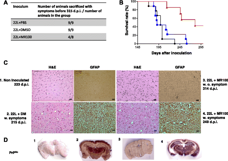
Pre-incubation of brain inoculum with MR100 substantially increases the survival time of injected animals. a Table with the number of animals sacrificed with symptoms before 315 days post-inoculation compared to the total number of animals for each group. b Kaplan-Meier survival plots of mice inoculated (15 μL/each mouse) with a 22 L-infected brain homogenate that was previously diluted in 2 % Sarkosyl/PBS and incubated at a final concentration of 1.5 mM MR100 (n = 8; 22 L + MR100; red) or an equivalent volume of DMSO (150 μL) (n = 10; 22 L + DM; blue) or PBS (150 μL) (n = 10; 22 L+ PBS; black) at room temperature for 2 h. c Histological analyses of thalamus (Th) sections from mice not inoculated with prions (1), or that were inoculated with 22 L + DM inoculum (2) or 22 L + MR100 inoculum and presented no symptoms (3) or with symptoms (4) of prion disease when they were killed (d.p.i., days post-inoculation). Tissue sections were stained with hematoxylin and eosin (HE) to confirm the prion pathology as indicated by the presence of vacuoles and probed with anti-GFAP antibodies as a marker of astrocytic gliosis. Tissue labeling was performed on several animals (2-3 per group) and images are representative of the staining observed in each group. d PET-blot analysis of frontal tissue sections of mice non-inoculated with prions (1), or that were inoculated with 22 L + DM inoculum (2), or 22 L + MR100 inoculum and presented no symptoms (3) or with symptoms (4) of prion disease when they were killed. The SAF84 antibody was used to detect rPrPSc proteins and the Vectastain ABC-AmP Kit (Vector laboratories, USA) to reveal antibody binding. Scale bars, 10 μM, w: with, w.o.: without symptoms

Similar articles

Cited by

References

    1. Prusiner SB. Novel proteinaceous infectious particles cause scrapie. Science. 1982;216(4542):136–144. doi: 10.1126/science.6801762. - DOI - PubMed
    1. Kovacs GG, Budka H. Molecular pathology of human prion diseases. Int J Mol Sci. 2009;10(3):976–999. doi: 10.3390/ijms10030976. - DOI - PMC - PubMed
    1. Caughey BW, Dong A, Bhat KS, Ernst D, Hayes SF, Caughey WS. Secondary structure analysis of the scrapie-associated protein PrP 27-30 in water by infrared spectroscopy. Biochemistry. 1991;30(31):7672–7680. doi: 10.1021/bi00245a003. - DOI - PubMed
    1. Pan KM, Baldwin M, Nguyen J, Gasset M, Serban A, Groth D, et al. Conversion of alpha-helices into beta-sheets features in the formation of the scrapie prion proteins. Proc Natl Acad Sci U S A. 1993;90(23):10962–10966. doi: 10.1073/pnas.90.23.10962. - DOI - PMC - PubMed
    1. Gambetti P, Dong Z, Yuan J, Xiao X, Zheng M, Alshekhlee A, et al. A novel human disease with abnormal prion protein sensitive to protease. Ann Neurol. 2008;63(6):697–708. doi: 10.1002/ana.21420. - DOI - PMC - PubMed

Publication types