. 2016 Mar 24;84(4):1062-1072.
doi: 10.1128/IAI.00843-15. Print 2016 Apr.

Uncovering an Important Role for YopJ in the Inhibition of Caspase-1 in Activated Macrophages and Promoting Yersinia pseudotuberculosis Virulence

Affiliations

Uncovering an Important Role for YopJ in the Inhibition of Caspase-1 in Activated Macrophages and Promoting Yersinia pseudotuberculosis Virulence

Taylor J Schoberle et al. Infect Immun. .

Abstract

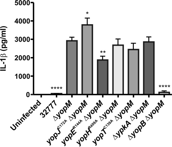
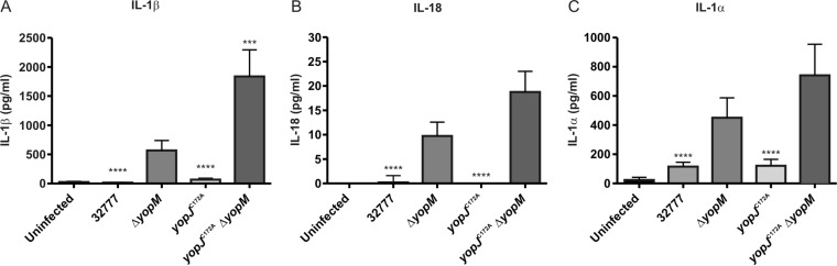
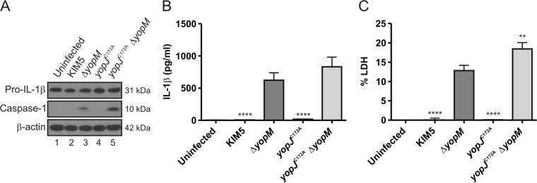
Pathogenic Yersinia species utilize a type III secretion system to translocate Yop effectors into infected host cells. Yop effectors inhibit innate immune responses in infected macrophages to promote Yersinia pathogenesis. In turn,Yersinia-infected macrophages respond to translocation of Yops by activating caspase-1, but different mechanisms of caspase-1 activation occur, depending on the bacterial genotype and the state of phagocyte activation. In macrophages activated with lipopolysaccharide (LPS) prior to Yersinia pseudotuberculosis infection, caspase-1 is activated by a rapid inflammasome-dependent mechanism that is inhibited by translocated YopM. The possibility that other effectors cooperate with YopM to inhibit caspase-1 activation in LPS-activated macrophages has not been investigated. Toward this aim, epistasis analysis was carried out in which the phenotype of aY. pseudotuberculosis yopM mutant was compared to that of a yopJ yopM, yopE yopM, yopH yopM, yopT yopM, or ypkA yopM mutant. Activation of caspase-1 was measured by cleavage of the enzyme, release of interleukin-1β (IL-1β), and pyroptosis in LPS-activated macrophages infected with wild-type or mutant Y. pseudotuberculosis strains. Results show enhanced activation of caspase-1 after infection with the yopJ yopM mutant relative to infection by any other single or double mutant. Similar results were obtained with the yopJ, yopM, and yopJ yopM mutants ofY ersinia pestis Following intravenous infection of mice, theY. pseudotuberculosis yopJ mutant was as virulent as the wild type, while the yopJ yopM mutant was significantly more attenuated than the yopM mutant. In summary, through epistasis analysis this work uncovered an important role for YopJ in inhibiting caspase-1 in activated macrophages and in promoting Yersinia virulence.


Figures

FIG 1
FIG 1
Measurement of IL-1β in supernatants of BMDMs infected with Y. pseudotuberculosis. BMDMs from C57BL/6 mice were primed with 100 ng/ml LPS for 18 h and left uninfected or infected with strain 32777 (wild type), a yopM mutant (ΔyopM), or yopJC172AΔyopM, yopER144AΔyopM, yopHR409AΔyopM, yopTC139A ΔyopM, ΔypkA ΔyopM, or ΔyopB ΔyopM double mutant at an MOI of 30. Concentrations of IL-1β in supernatants collected at 90 min postinfection were quantified by ELISA. Data represent average values ± the standard errors of the means from three independent experiments. *, P < 0.05; **, P < 0.01; ****, P < 0.0001, as determined by one-way analysis of variance compared to results for ΔyopM strain-infected macrophages.
FIG 2
FIG 2
Measurement of caspase-1-dependent proinflammatory cytokines in supernatants of BMDMs infected with Y. pseudotuberculosis. BMDMs from C57BL/6 mice were primed with 100 ng/ml LPS for 18 h and left uninfected or infected with the 32777, ΔyopM, yopJC172A, or yopJC172A ΔyopM strain at an MOI of 30. At 90 min postinfection, supernatants were collected, and concentrations of secreted IL-1β, IL-18, or IL-1α were measured by ELISA. Data represent average values ± the standard errors of the means from three independent experiments. ***, P < 0.001; ****, P < 0.0001, as determined by one-way analysis of variance compared to values for 32777 ΔyopM-infected macrophages.
FIG 3
FIG 3
Measurement of processed caspase-1, pro-IL-1β, mature IL-1β, and pyroptosis in BMDMs infected with Y. pseudotuberculosis. LPS-primed BMDMs from C57BL/6 mice were left uninfected or infected with the 32777, ΔyopM, or yopJC172AΔyopM strain at an MOI of 30. (A) After a 90-min infection, lysates were prepared and subjected to Western blotting with antibodies recognizing pro-IL-1β, caspase-1, or β-actin. Molecular masses of corresponding proteins are shown on the right. Quantification of the processed caspase-1 bands showed that the signal for the yopJC172AΔyopM strain was 4.7 times greater than that for the ΔyopM strain. Supernatants were harvested at 90 min postinfection, and secreted IL-1β was measured by ELISA (B); pyroptosis was measured by quantification of percent LDH release (C). The data in panels B and C represent average values ± the standard errors of the means from seven independent experiments. ***, P < 0.001; ****, P < 0.0001, as determined by one-way analysis of variance compared to results for ΔyopM mutant-infected macrophages.
FIG 4
FIG 4
Determination of survival and organ colonization of mice infected with Y. pseudotuberculosis. C57BL/6 mice were infected i.v. with ∼1,000 CFU of the indicated Y. pseudotuberculosis strain. (A) Time to death was monitored for 14 days [n = 14 for 32777 and the yopJC172AΔyopM strains; n = 10 for the ΔyopM and yopJC172A strains; and n = 8 for the yopJC172AΔyopM(pYopM) strain]. (B and C) At 4 days postinfection, mice were euthanized, and organs were collected and processed for CFU assay (n = 11 for 32777 and the ΔyopM strains; n = 14 for the yopJC172A strain; and n = 17 for yopJC172AΔyopM strain). Data from all spleens and the subset of livers analyzed are shown, with horizontal bars indicating arithmetic means. Data in panels A, B, and C represent values from four or five independent experiments. (D and E) At 6 days postinfection, mice were euthanized, and organs were collected and processed for CFU assay (n = 8 for the ΔyopM strain, and n = 8 for the yopJC172AΔyopM strain). Data in panels D and E represent values from two independent experiments. Significance of differences between survival curves in panel A was determined by log rank test, while the significance of differences in organ colonization in panels B and C was calculated using a Mann-Whitney test, in all cases compared to values for ΔyopM mutant-infected mice. *, P < 0.05; **, P < 0.01; ****, P < 0.0001. All CFU values were analyzed by Grubb's test to identify significant outliers, and all data from any mouse found to have an outlying value are not shown and were removed prior to the calculation of significance.
FIG 5
FIG 5
Measurement of processed caspase-1, pro-IL-1β, mature IL-1β, and pyroptosis in BMDMs infected with Y. pseudotuberculosis or Y. pestis. LPS-primed BMDMs were left uninfected or infected with 32777, 32777 ΔyopM, KIM5, or KIM5 ΔyopM for 90 min at an MOI of 30. (A) After infection, lysates were collected, processed, and subjected to Western blotting using antibodies to pro-IL-1β, cleaved caspase-1, or β-actin. Supernatants were collected and analyzed for secreted IL-1β (B) or pyroptosis by measurement of LDH release (C). Data represent average values ± the standard errors of the means from three independent experiments. ****, P < 0.0001, as determined by one-way analysis of variance comparing values in BMDMs infected with 32777 ΔyopM versus those with 32777 infection or BMDMs infected with KIM5 ΔyopM versus those infected with KIM5.
FIG 6
FIG 6
Measurement of processed caspase-1, pro-IL-1β, mature IL-1β, and pyroptosis in BMDMs infected with Y. pestis. BMDMs from C57BL/6 mice were primed with 100 ng/ml LPS for 18 h and left uninfected or infected with KIM5, KIM5 ΔyopM, KIM5 yopJC172A, or KIM5 yopJC172AΔyopM at an MOI of 30. (A) At 90 min postinfection, lysates were collected, processed, and subjected to Western blotting using antibodies against pro-IL-1β and p10, the 10-kDa subunit of caspase-1. β-Actin was blotted as a loading control. Quantification of the processed caspase-1 bands showed that the signal for KIM5 yopJC172AΔyopM was 2.3 times greater than that for KIM5 ΔyopM. At 90 min postinfection, supernatants were collected, and secreted IL-1β was measured by ELISA (B); pyroptosis was quantified as percent LDH release (C). The data in panel B and C represent average values ± the standard error of the means from three independent experiments. **, P < 0.01; ****, P < 0.0001, as determined by one-way analysis of variance compared to results for KIM5 ΔyopM-infected macrophages.
FIG 7
FIG 7
Measurement of processed caspase-1, pro-IL-1β, mature IL-1β, and pyroptosis in BMDMs infected with Y. pestis strains expressing different YopJ isoforms. LPS-primed BMDMs from C57BL/6 mice were left uninfected or infected with KIM5 ΔyopM strains expressing yopJKIM, yopJYPTB, yopJCO92, or yopJC172A at an MOI of 30. (A) Lysates were collected and analyzed at 90 min postinfection using antibodies against pro-IL-1β, cleaved caspase-1, or β-actin. Quantification of the processed caspase-1 bands showed that the signal for KIM5 yopJC172A ΔyopM was approximately 1.4 times greater than that for KIM5 ΔyopM expressing the different isoforms. Supernatants were collected at 90 min postinfection and analyzed for secreted IL-1β by ELISA (B); pyroptosis was quantified by measuring LDH release (C). Data represent average values ± the standard errors of the means from three independent experiments. By one-way analysis of variance there were no significant differences between results for the KIM5 yopJKIM ΔyopM strain and those for the other strains tested.

Similar articles

Cited by

References

    1. Medzhitov R. 2007. Recognition of microorganisms and activation of the immune response. Nature 449:819–826. doi:10.1038/nature06246. - DOI - PubMed
    1. Motta V, Soares F, Sun T, Philpott DJ. 2015. NOD-like receptors: versatile cytosolic sentinels. Physiol Rev 95:149–178. doi:10.1152/physrev.00009.2014. - DOI - PubMed
    1. Lawrence T. 2009. The nuclear factor NF-κB pathway in inflammation. Cold Spring Harb Perspect Biol 1:a001651. doi:10.1101/cshperspect.a001651. - DOI - PMC - PubMed
    1. Kawai T, Akira S. 2007. Signaling to NF-κB by Toll-like receptors. Trends Mol Med 13:460–469. doi:10.1016/j.molmed.2007.09.002. - DOI - PubMed
    1. Finlay BB, McFadden G. 2006. Anti-immunology: evasion of the host immune system by bacterial and viral pathogens. Cell 124:767–782. doi:10.1016/j.cell.2006.01.034. - DOI - PubMed

Publication types

MeSH terms