. 2016 Apr;131(4):571-85.
doi: 10.1007/s00401-016-1537-1. Epub 2016 Jan 25.

Updated TDP-43 in Alzheimer's disease staging scheme

Affiliations

Updated TDP-43 in Alzheimer's disease staging scheme

Keith A Josephs et al. Acta Neuropathol. 2016 Apr.

Abstract

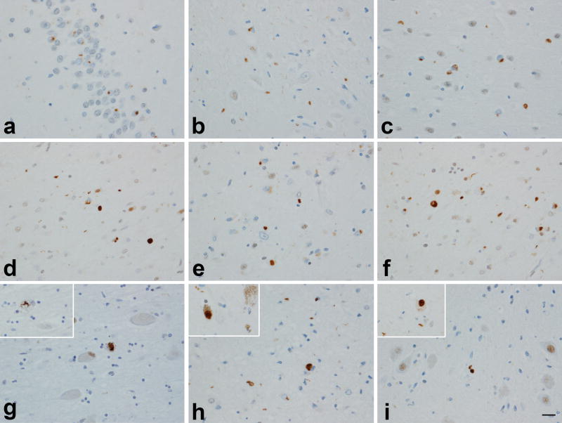
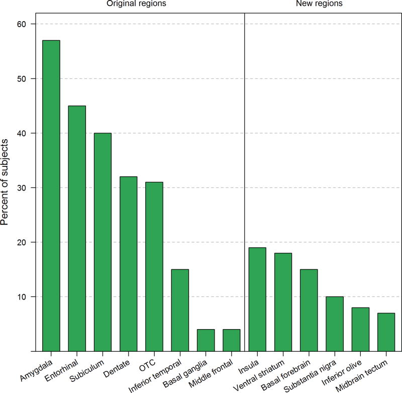
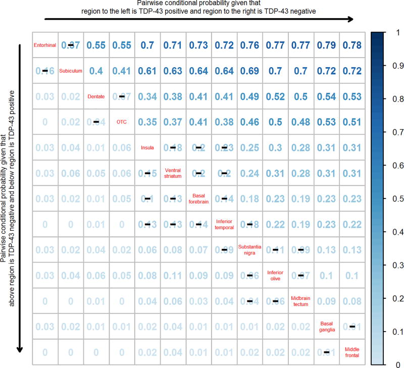
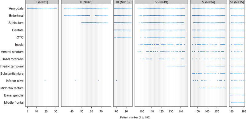
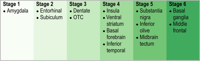
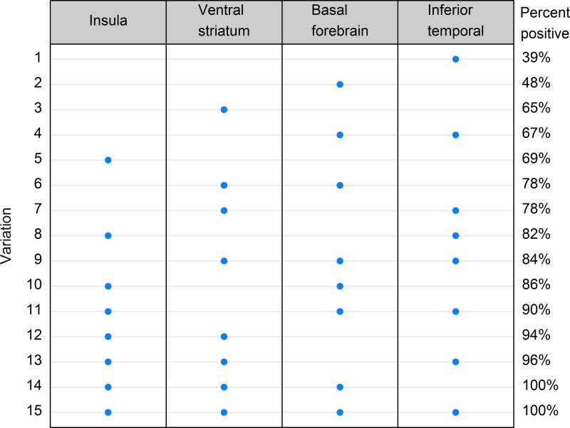
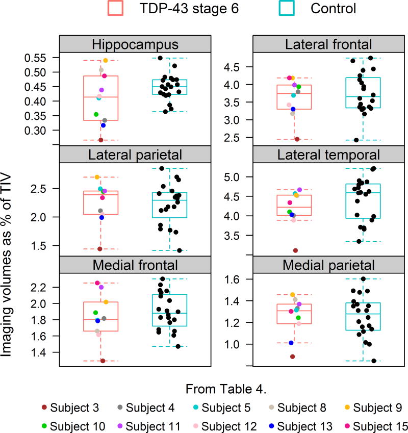
In this study, we update the TDP-43 in Alzheimer's disease staging scheme by assessing the topography of TDP-43 in 193 cases of Alzheimer's disease, in 14 different brain regions (eight previously described plus six newly reported) and use conditional probability to model the spread of TDP-43 across the 14 brain regions. We show that in addition to the eight original regions we previously reported [amygdala, entorhinal cortex, subiculum, dentate gyrus of the hippocampus, occipitotemporal cortex, inferior temporal cortex, middle frontal cortex and basal ganglia (putamen/globus pallidum)] that TDP-43 is also deposited in the insular cortex, ventral striatum, basal forebrain, substantia nigra, midbrain tectum, and the inferior olive of the medulla oblongata, in Alzheimer's disease. The conditional probability analysis produced six significantly different stages (P < 0.01), and suggests that TDP-43 deposition begins in the amygdala (stage 1), then moves to entorhinal cortex and subiculum (stage 2); to the dentate gyrus of the hippocampus and occipitotemporal cortex (stage 3); insular cortex, ventral striatum, basal forebrain and inferior temporal cortex (stage 4); substantia nigra, inferior olive and midbrain tectum (stage 5); and finally to basal ganglia and middle frontal cortex (stage 6). This updated staging scheme is superior to our previous staging scheme, classifying 100% of the cases (versus 94% in the old scheme), based on criteria provided, and shows clinical significance with some regions and with increasing stage. We discuss the relevance of the updated staging scheme, as well as its impact on the prion-like hypothesis of protein spread in neurodegenerative disease. We also address the issue of whether frontotemporal lobar degeneration with TDP-43 could be the primary pathology in stage 6.

Keywords: Alzheimer’s disease; Brainstem; Insular cortex; Limbic; Staging; TDP-43.


Figures

Figure 1
Figure 1
TDP-43 deposition across different regions in cases with high probability Alzheimer’s disease: dentate fascia (a); subiculum (b); entorhinal cortex (c); amygdala (d); ventral striatum (e); insular cortex (f); basal nucleus (inset: NCI) (g); midbrain tectum (inset: substantia nigra) (h); medulla – inferior olivary nucleus (inset: NCI) (i). In most instances TDP-43 immunoreactive neuronal cytoplasmic inclusions were observed although dystrophic neurites can also be seen in many panels. Magnification × 200 (inset × 400).
Figure 2
Figure 2
Bar plot showing the frequency of TDP-43 deposition in the eight original and six newly assessed regions among 340 cases. The most common affected region was the amygdala (frequency=56%).
Figure 3
Figure 3
A pairwise conditional probability matrix of the regions analyzed. Reading the plot from left to right, the conditional probability estimates show the estimated probability that the region on the left is TDP-43 positive before the region on the right. For example, the probability of entorhinal being TDP-43 positive given that subiculum is TDP-43 negative is 0.37. Reading the plot from top to bottom, the entries show the estimated probability that the region below is TDP-43 positive before the region above. For example, the probability of subiculum being TDP-43 positive given that entorhinal is negative is 0.16. Black lines (–) across conditional probability estimates indicate p-values are not statistically significant at the < 0.01 level. Note however, p-values between inferior temporal cortex and substantia nigra (p=0.01), between (insula, ventral striatum and basal forebrain) and substantia nigra (p<0.001), and between (insula, ventral striatum, basal forebrain, inferior temporal cortex) and (inferior olive and midbrain tectum (P<0.001). P-values were assessed using exact McNemar’s test.
Figure 4
Figure 4
Patterns of TDP-43 positivity across 14 regions for 193 cases. The vertical axis indicates regions and the horizontal axis indicates patients. A blue dot indicates the case was TDP-43 positive for that region. Patients are grouped by TDP-43 in Alzheimer’s disease stage.
Figure 5
Figure 5
Diagram illustrating the TDP-43 in Alzheimer’s disease stage progression.
Figure 6
Figure 6
A plot of percent of stage 4 cases with TDP-43 deposition in 15 combinations of all four regions (insula cortex, ventral striatum, basal forebrain and inferior temporal cortex). The plot is ordered by percent TDP-43 positive from smallest to largest.
Figure 7
Figure 7
A comparison of regional volumes between 10 stage 6 cases that had antemortem volumetric head MRI and 20 age, gender, and NFT Braak stage matched Alzheimer’s disease cases without TDP-43.

Similar articles

Cited by

References

    1. A R. L'examen clinique en psychologie. Presses Universitaires de France; City: 1964.
    1. Aguzzi A, Rajendran L. The transcellular spread of cytosolic amyloids, prions, and prionoids. Neuron. 2009;64:783–790. doi: 10.1016/j.neuron.2009.12.016. - DOI - PubMed
    1. Amador-Ortiz C, Lin WL, Ahmed Z, et al. TDP-43 immunoreactivity in hippocampal sclerosis and Alzheimer's disease. Ann Neurol. 2007;61:435–445. doi: 10.1002/ana.21154. - DOI - PMC - PubMed
    1. Arai T, Hasegawa M, Akiyama H, et al. TDP-43 is a component of ubiquitin-positive tau-negative inclusions in frontotemporal lobar degeneration and amyotrophic lateral sclerosis. Biochem Biophys Res Commun. 2006;351:602–611. doi: 10.1016/j.bbrc.2006.10.093. - DOI - PubMed
    1. Arai T, Mackenzie IR, Hasegawa M, et al. Phosphorylated TDP-43 in Alzheimer's disease and dementia with Lewy bodies. Acta Neuropathol. 2009;117:125–136. doi: 10.1007/s00401-008-0480-1. - DOI - PubMed

Publication types

LinkOut - more resources