Clinical Trial
. 2016 Mar 1;196(5):2167-80.
doi: 10.4049/jimmunol.1501853. Epub 2016 Jan 25.

Yeast-Derived Particulate β-Glucan Treatment Subverts the Suppression of Myeloid-Derived Suppressor Cells (MDSC) by Inducing Polymorphonuclear MDSC Apoptosis and Monocytic MDSC Differentiation to APC in Cancer

Affiliations
Clinical Trial

Yeast-Derived Particulate β-Glucan Treatment Subverts the Suppression of Myeloid-Derived Suppressor Cells (MDSC) by Inducing Polymorphonuclear MDSC Apoptosis and Monocytic MDSC Differentiation to APC in Cancer

Sabrin H Albeituni et al. J Immunol. .

Erratum in

Abstract

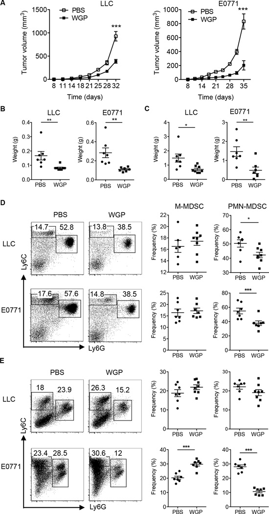
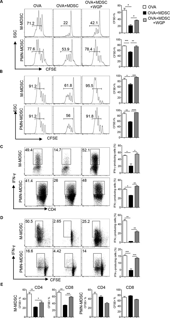
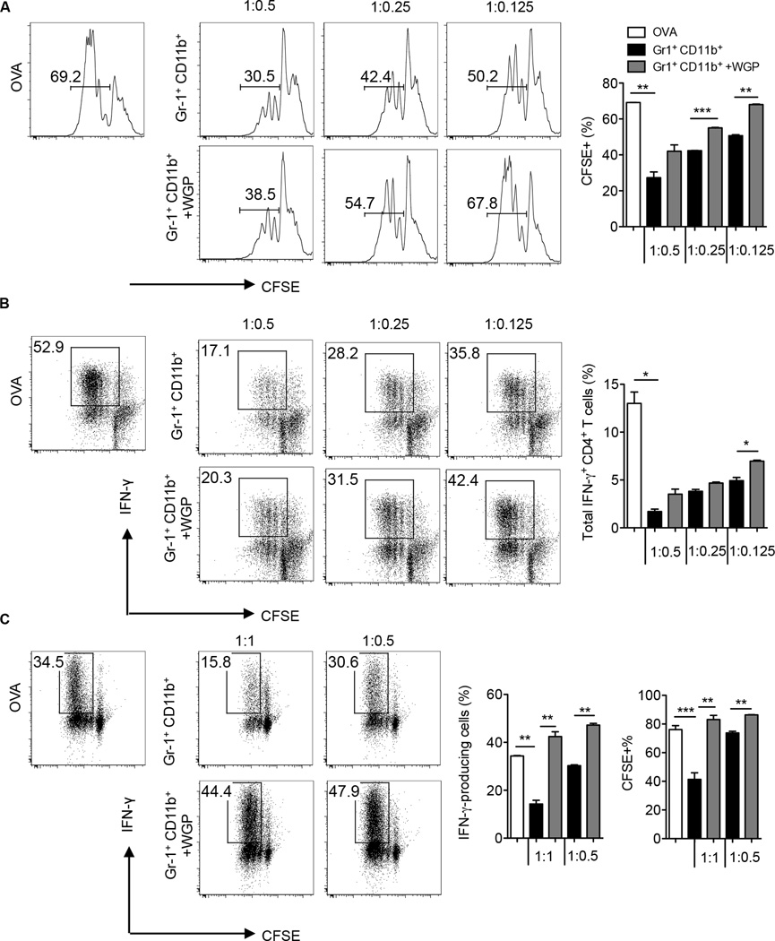
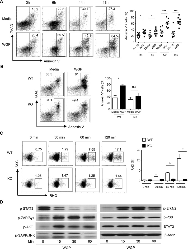
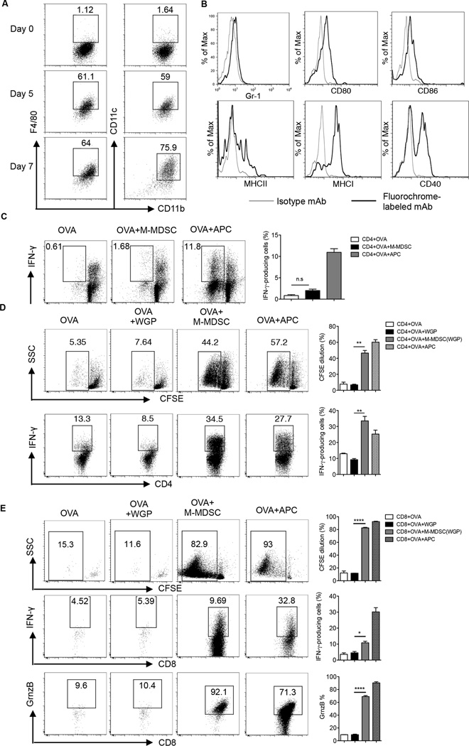
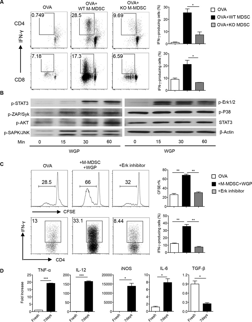
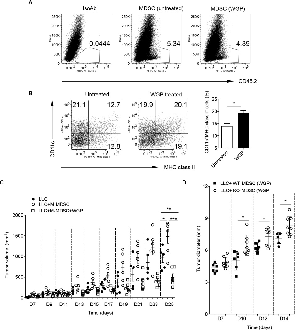
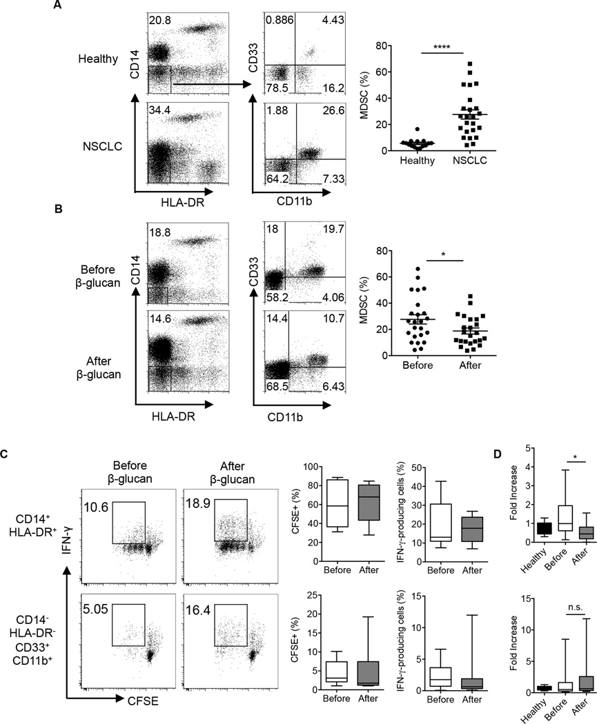
Myeloid-derived suppressor cells (MDSC) are a heterogeneous population of immature myeloid cells that promote tumor progression. In this study, we demonstrated that activation of a C-type lectin receptor, dectin-1, in MDSC differentially modulates the function of different MDSC subsets. Yeast-derived whole β-glucan particles (WGP; a ligand to engage and activate dectin-1, oral treatment in vivo) significantly decreased tumor weight and splenomegaly in tumor-bearing mice with reduced accumulation of polymorphonuclear MDSC but not monocytic MDSC (M-MDSC), and decreased polymorphonuclear MDSC suppression in vitro through the induction of respiratory burst and apoptosis. On a different axis, WGP-treated M-MDSC differentiated into F4/80(+)CD11c(+) cells in vitro that served as potent APC to induce Ag-specific CD4(+) and CD8(+) T cell responses in a dectin-1-dependent manner. Additionally, Erk1/2 phosphorylation was required for the acquisition of APC properties in M-MDSC. Moreover, WGP-treated M-MDSC differentiated into CD11c(+) cells in vivo with high MHC class II expression and induced decreased tumor burden when inoculated s.c. with Lewis lung carcinoma cells. This effect was dependent on the dectin-1 receptor. Strikingly, patients with non-small cell lung carcinoma that had received WGP treatment for 10-14 d prior to any other treatment had a decreased frequency of CD14(-)HLA-DR(-)CD11b(+)CD33(+) MDSC in the peripheral blood. Overall, these data indicate that WGP may be a potent immune modulator of MDSC suppressive function and differentiation in cancer.


Figures

Figure 1
Figure 1. Particulate β-glucan treatment in vivo reduces tumor burden and impacts the frequency of MDSC in spleens and tumors of LLC and E0771-bearing mice
(A) C57BL/6 WT mice (n=7, 8) were injected subcutaneously (s.c) with LLC or E0771 tumor cell lines. Once palpable tumors were formed (day 8), mice were orally administered with particulate β-glucan (800 µg, daily) or PBS with a gavage needle at indicated time. Tumor diameters were measured every three days and tumor volumes were then calculated. (B) On day 32 (LLC model) or day 35 (E0771 model), mice were killed and spleens were excised and weighed. Each point in the data plot represents the spleen weight of each mouse in grams. PBS-treated group was compared to particulate β-glucan treated group (WGP) in both models (C) Tumor tissues were excised and weighted from WGP or PBS-treated mice. (D) Flow cytometry analysis of the frequencies of M-MDSC (Ly6GLy6Chigh) and PMN-MDSC (Ly6G+Ly6Cint) in the spleens of LLC and E0771-bearing mice treated with PBS or particulate β-glucan (WGP). Cells were gated on CD11b+ cells. (E) Frequencies of M-MDSC and PMN-MDSC in the tumors of LLC and E0771-bearing mice treated with PBS or particulate β-glucan (WGP). Cells were gated on CD45+CD11b+ cells. *p<0.05, ** p<0.01, *** p<0.001.
Figure 2
Figure 2. Particulate β-glucan treatment in vitro subverts splenic MDSC-mediated T cell suppression
(A) CFSE-labeled OT-II splenocytes co-cultured with sorted M-MDSC or PMN-MDSC from the spleens of LLC-bearing mice in the presence or absence of particulate β-glucan (100 µg/ml in the M-MDSC cultures and 50 µg/ml in the PMN-MDSC cultures) and OVA (100 µg/ml) for 3 days at 1:1 ratio. Data represent the percentage of CFSE diluted cells gated on CD4+ T cells. The experiment was repeated two times with similar results. (B) CFSE-labeled OT-I splenocytes were co-cultured with sorted M-MDSC or PMN-MDSC from the spleens of LLC-bearing mice in the presence of OVA (50 µg/ml) and particulate β-glucan (50 µg/ml) for 3 days at 1:1 ratio. Data represent the percentage of CFSE diluted cells gated on CD8+ T cells. Data is representative of three independent experiments. (C) OT-II splenocytes co-cultured with sorted M-MDSC or PMN-MDSC from the spleens of LLC-bearing mice in the presence of OVA (100 µg/ml) with or without particulate β-glucan (50 µg/ml) for 3–4 days at 1:1 ratio and stimulated with PMA/Ionomycin for intracellular IFN-γ staining. Data represent the percentage of IFN-γ+ cells gated on CD4+ T cells. The experiment was repeated 3 times with similar results. (D) CFSE-labeled OT-I splenocytes co-cultured with sorted M-MDSC or PMN-MDSC from the spleens of LLC-bearing mice in the presence of OVA (50 µg/ml in M-MDSC cultures and 10 µg/ml in PMN-MDSC cultures) and particulate β-glucan (50 µg/ml) for 3 days at 1:1 ratio and then stimulated with PMA/Ionomycin for intracellular IFN-γ staining. Data represent the percentage of IFN-γ+ CFSE diluted cells gated on CD8+ T cells. Results are representative of three independent experiments. (E) CFSE-labeled splenocytes from C57BL/6 mice stimulated with plate-bound anti-CD3 (5 µg/ml) and soluble anti-CD28 (2 µg/ml) (white bar) and co-cultured with sorted splenic M-MDSC or PMN-MDSC from LLC-bearing mice for 3 days with (black bar) or without particulate β-glucan (50 µg/ml) (grey bar). Data represent the frequency of CFSE diluted cells gated on CD4+ or CD8+ T cells. The experiment was repeated twice with similar results. * p<0.05, **p<0.01, ***p<0.001.
Figure 3
Figure 3. Particulate β-glucan reduces tumor Gr-1+CD11b+ MDSC-mediated T cell suppression
(A) Tumor Gr-1+CD11b+CD45+ MDSC sorted from LLC-bearing mice were co-cultured with CFSE-labeled OT-II splenocytes at indicated ratios, in the presence of OVA (100 µg/ml) with or without particulate β-glucan (50 µg/ml) for 3–4 days. Data represent the frequency of CFSE diluted cells gated on CD4+ T cells. The experiment was repeated twice with similar results. (B) Same cell cultures as (A) were further stimulated with PMA/Ionomycin for intracellular IFN-γ staining. The experiment was repeated twice with similar results. (C) Splenocytes of OT-I mice were co-cultured with sorted Gr-1+CD11b+CD45+ tumor MDSC from LLC-bearing mice at indicated ratios, in the presence of OVA (50 µg/ml in M-MDSC cultures and 10 µg/ml in PMN-MDSC cultures) with or without particulate β-glucan (50 µg/ml). Data represent the percentage of IFN-γ+ cells and CFSE diluted cells gated on CD8+ T cells. Results are representative of two independent experiments. *p<0.05, **p<0.01, ***p<0.001.
Figure 4
Figure 4. Particulate β-glucan induces respiratory burst and apoptosis in PMN-MDSC in a dectin-1 dependent manner
(A) Representative dot plots showing the frequency of annexin V+ cells gated on Gr-1highCD11b+ PMN-MDSC in splenocytes of LLC-bearing mice cultured with media only or with particulate β-glucan (100 µg/ml) for indicated time. Summarized data are also shown (n=8). (B) Frequencies of annexinV+ cells gated on Gr-1highCD11b+ PMN-MDSC from WT or dectin-1 KO LLC-bearing mice cultured for 18 hours with or without particulate β-glucan (100 µg/ml) (n=2). (C) Respiratory burst in PMN-MDSC sorted from spleens of LLC-bearing WT or dectin-1 KO mice stimulated with particulate β-glucan (100 µg/ml) for indicated time. Reduction of dihydrorhodamine 123 (DHR) to fluorescent rhodamine 123 (RHO) was assessed by flow cytometry. Data is representative of 6 independent experiments (WT) and two independent experiments (KO). (D) Western blot analysis of p-STAT3, p-Zap/Syk, p-Akt, p-SAPK/JNK, p-Erk1/2, p-p38, STAT3 and β-actin in PMN-MDSC sorted from the spleens of LLC-bearing mice and treated with particulate β-glucan (100 µg/ml) for 0, 15, 30 and 60 minutes. Results are representative of at least three independent experiments. *p<0.05, **p<0.01, ***p<0.001.
Figure 5
Figure 5. Particulate β-glucan induces the differentiation of M-MDSC to antigen-presenting cells in vitro
(A) Expression of CD11c, F4/80 and CD11b surface markers on M-MDSC cultured with particulate β-glucan (50 µg/ml) for 0, 5 and 7 days. Results were repeated at least three times with similar results. (B) Expression of Gr-1, CD80, CD86, MHC class II, MHC class I, and CD40 surface markers on M-MDSC cultured with particulate β-glucan (50 µg/ml) for 7 days. Histograms represent the results of three independent experiments. (C) Frequency of IFN-γ+ CFSE gated on CD4+ T cells in cultures where freshly sorted M-MDSC from the spleens of LLC-bearing mice were incubated for 5 days with CFSE-labeled CD4+ T cells in the presence of OVA (100µg/ml). Irradiated splenocytes were used as APC positive control. (D) Sorted and CFSE-labeled CD4+ OT-II T cells were co-cultured with splenic M-MDSC pre-cultured with particulate β-glucan for 7 days in the presence of OVA (50 µg/ml) for 4–5 days. The frequencies of CFSE diluted cells and IFN-γ+ CD4+ T cells were shown. Irradiated splenocytes were used as APC positive control. The results are representative of at least four independent experiments. (E) Sorted and CFSE-labeled CD8+ OT-I T cells were co-cultured for 4–5 days with OVA (50 µg/ml) and splenic M-MDSC pre-cultured with particulate β-glucan for 7 days. The frequencies of CFSE diluted cells, IFN-γ+ cells and Granzyme B+ cells gated on CD8+ T cells are demonstrated. Results are representative of two independent experiments. *p<0.05, **p<0.01, ***p<0.001, ****p<0.0001.
Figure 6
Figure 6. Particulate β-glucan-induced M-MDSC antigen-presenting function is dectin-1 and Erk1/2 dependent
(A) CD4+ and CD8+ T cells were sorted from OT-II and OT-I mice, respectively, CFSE-labeled and co-cultured for 4–5 days with M-MDSC sorted from the spleens of WT or dectin-1 KO LLC-bearing mice. M-MDSC were pre-treated with particulate β-glucan for 7 days prior to co-culture with CD4+ or CD8+ T cells. The frequency of CFSEIFN-γ+, gated on CD4+ T cells (upper panel) or CD8+ T cells (lower panel), are represented in the dot plots. Results are representative of two-independent experiments. (B) Western blot analysis of p-STAT3, p-Zap/Syk, p-Akt, p-SAPK/JNK, p-Erk1/2, p-p38, STAT3 and β-actin in M-MDSC sorted from the spleens of LLC-bearing mice and stimulated with particulate β-glucan (100 µg/ml) for 0, 15, 30 and 60 minutes. (C) CD4+ T cells sorted from OT-II mice were CFSE-labeled and co-cultured with OVA and M-MDSC pre-treated with particulate β-glucan in the presence of MEK1/2 inhibitor (PD98059) (30 ng/ml) or DMSO for 7 days. Data demonstrate the frequencies of CFSE diluted cells and IFN-γ+ cells gated on CD4+ T cells. Results are representative of two independent experiments. (D) The relative mRNA expression of TNF-α, IL-12, iNOS, IL-6, and TGF-β in MDSC, sorted from the spleens of LLC-bearing mice, and incubated with particulate β-glucan for 7 days compared to its expression in freshly sorted M-MDSC. *p<0.05, **p<0.01.
Figure 7
Figure 7. Particulate β-glucan treatment induces M-MDSC differentiation in vivo with reduced tumor growth
Tumor M-MDSC sorted from C57Bl/6 LLC tumor-bearing mice (CD45.2) were treated with or without WGP for overnight and then intratumorally injected into SJL tumor-bearing mice (CD45.1). Mice were sacrificed after 7 days and single cell suspensions from tumors were stained with anti-CD45.2 or isotype control mAb (A) and CD11c and MHC class II. Cells were gated on CD45.2+ cells (B). The percentage of CD11c+MHC class II+ cells was summarized. (C) M-MDSC sorted from LLC tumor-bearing mice were treated with or without WGP and then mixed with LLC cells for injection. LLC alone was used as control. Tumor growth was monitored and recorded. (D) M-MDSC sorted from LLC tumor-bearing wildtype or dectin-1 KO mice were treated with WGP and mixed with LLC cells for injection. Tumor growth was monitored. *p<0.05, **p<0.01, ***p<0.001.
Figure 8
Figure 8. Particulate β-glucan treatment in vivo decreased the frequency of CD14HLA-DRCD33+CD11b+ MDSC in the peripheral blood of NSCLC patients
(A) Frequency of CD33+CD11b+ MDSC gated on CD14HLA-DR cells in the peripheral blood of NSCLC patients (n=23) compared to age and sex matched healthy donors (n=13). (B) Frequency of CD33+CD11b+ MDSC gated on CD14HLA-DR cells in the peripheral blood of NSCLC patients before and after particulate β-glucan treatment for 10–14 days (n=23). (C) IFN-γ production and proliferation (CFSE diluted cells) of allogeneic T cells (CD3+) cultured at 1:1 ratio with CD14+HLA-DR+CD11b+CD33+ or CD14HLA-DRCD11b+CD33+ isolated from the peripheral blood of NSCLC patients before and after particulate β-glucan treatment (n=11) (D) Relative Arginase1 mRNA expression levels in neutrophils (PMN) isolated from the peripheral blood of healthy controls, and patients with NSCLC before and after particulate β-glucan treatment. Arginase 1 relative mRNA expression significantly decreased in a cohort of 15 patients, whereas no significant change was reported in the other 20 patients (total n=35). *p<0.05, **p<0.01, ***p<0.001.

Similar articles

Cited by

References

    1. Dunn GP, Bruce AT, Ikeda H, Old LJ, Schreiber RD. Cancer immunoediting: from immunosurveillance to tumor escape. Nat Immunol. 2002;3:991–998. - PubMed
    1. Dunn GP, Old LJ, Schreiber RD. The three Es of cancer immunoediting. Annu Rev Immunol. 2004;22:329–360. - PubMed
    1. Schreiber RD, Old LJ, Smyth MJ. Cancer immunoediting: integrating immunity’s roles in cancer suppression and promotion. Science. 2011;331:1565–1570. - PubMed
    1. Gabrilovich DI, Nagaraj S. Myeloid-derived suppressor cells as regulators of the immune system. Nat Rev Immunol. 2009;9:162–174. - PMC - PubMed
    1. Youn JI, Nagaraj S, Collazo M, Gabrilovich DI. Subsets of myeloid-derived suppressor cells in tumor-bearing mice. J Immunol. 2008;181:5791–5802. - PMC - PubMed

Publication types

MeSH terms