Case Reports
. 2016 Jan 25;14(1):22.
doi: 10.1186/s12957-016-0774-z.

Cytokeratin 7-positive/cytokeratin 20-negative cecal adenocarcinoma metastatic to the uterine cervix: a case report

Affiliations
Case Reports

Cytokeratin 7-positive/cytokeratin 20-negative cecal adenocarcinoma metastatic to the uterine cervix: a case report

Masafumi Toyoshima et al. World J Surg Oncol. .

Abstract

Background: The vast majority of uterine cervical malignancies are primary carcinomas, and secondary neoplasms that metastasize to the uterine cervix from a distant organ are uncommon. Although relatively rare, metastases to the uterine cervix from a primary colon cancer have been reported. We report a rare case of metastatic carcinoma originating from a cecal adenocarcinoma with an unusual cytokeratin 7/cytokeratin 20 immunophenotype.

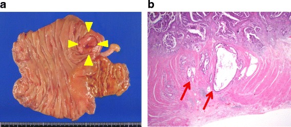
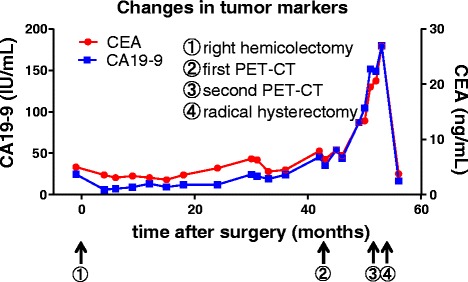
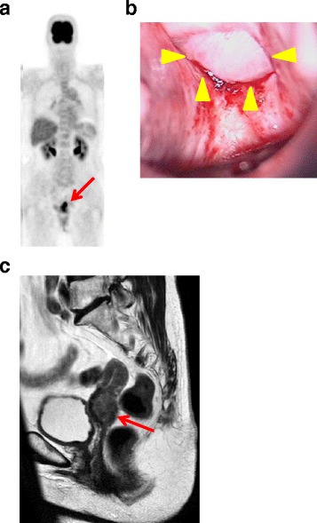
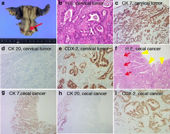
Case presentation: A 74-year-old postmenopausal Japanese woman was referred to our hospital for the evaluation of a uterine tumor. She had a past medical history of cecal cancer and had undergone laparoscopically assisted right hemicolectomy at the age of 69 years. During follow-up, she was found to have elevated levels of the tumor markers carbohydrate antigen 19-9 (179.7 IU/mL) and carcinoembryonic antigen (26.9 μg/L). Positron emission tomography/computed tomography showed a focus of high 18F-fluorodeoxyglucose uptake in her uterus. Examination of a cervical biopsy found a poorly differentiated adenocarcinoma that was immunopositive for cytokeratin (CK)7 and caudal-related homeobox 2 (CDX2) expression and immunonegative for cytokeratin 20 expression. The patient underwent radical hysterectomy and bilateral salpingo-oophorectomy. Histopathological examination found invasive growth of irregular and atypical ductal hyperplasia. Immunohistochemical staining of the tumor specimen revealed the same immunophenotype as the biopsy specimen. The cecal cancer specimen from her previous surgery was also examined and found to have the same immunophenotype. The histopathological diagnosis was cecal adenocarcinoma metastatic to the uterine cervix. The patient is currently receiving adjuvant chemotherapy and to date is without evidence of recurrent disease.

Conclusions: Our report illustrates the importance of immunohistochemistry for the correct diagnosis of the origin of a uterine cervical adenocarcinoma in a patient with a medical history of colorectal cancer. Re-examination of a previous oncological specimen is critical for cases with a uterine lesion that is difficult to identify as primary or metastatic cancer.


Figures

Fig. 1
Fig. 1
Pathology of the primary cecal carcinoma. a Gross appearance of the ulcerous tumor in the cecum (yellow arrowheads). Note that a normal appendix is apparent. b Hematoxylin-and-eosin-stained section of the primary cecal cancer specimen demonstrates that the adenocarcinoma has slightly invaded the muscle layer to reach the subserosa (red arrows) (original magnification ×100)
Fig. 2
Fig. 2
Timeline of clinical events and changes in CEA and CA19-9 levels
Fig. 3
Fig. 3
Imaging and physical examinations. a Positron emission tomography-computed tomography shows high 18F-fluorodeoxyglucose uptake consistent with a uterine tumor (red arrow). b Colposcopic view shows a solid white tumor on the posterior fornix (yellow arrowheads). c Pelvic magnetic resonance image of a T2-enhanced sagittal section showing an irregularly enlarged uterine cervical tumor (red arrow)
Fig. 4
Fig. 4
Gross appearance of resected uterus, histopathology, and immunohistochemistry of sections of uterine cervical tumor and cecal tumor specimens. a Gross appearance of the resected uterus. Note that the mucosal surface of the endocervical canal is clear (red arrow). b Hematoxylin and eosin staining of the cervical tumor specimen demonstrates invasive growth of irregular and atypical ductal hyperplasia with moderate differentiation (original magnification ×200). c Cervical carcinoma shows diffuse and strongly positive CK7 staining (original magnification ×100). d Cervical carcinoma is negative for CK20 staining (original magnification ×100). e Cervical carcinoma cells show diffuse and strongly positive CDX2 nuclear staining (original magnification ×200). f Hematoxylin-and-eosin-stained section of the cecal adenocarcinoma demonstrates well-differentiated adenocarcinoma (yellow arrowheads). Note that normal crypt architecture is seen on the left side of the picture (red arrows) (original magnification ×100). g Cecal carcinoma shows diffuse and strongly positive CK7 staining (original magnification ×100). h Cecal carcinoma is negative for CK20 staining. Note that normal crypt architecture is positive for CK20 staining (original magnification ×100). i Cecal carcinoma shows diffuse and strongly positive CDX2 nuclear staining (original magnification ×200)

Similar articles

Cited by

References

    1. Lemoine NR, Hall PA. Epithelial tumors metastatic to the uterine cervix. A study of 33 cases and review of the literature. Cancer. 1986;57:2002–5. doi: 10.1002/1097-0142(19860515)57:10<2002::AID-CNCR2820571021>3.0.CO;2-L. - DOI - PubMed
    1. Nakagami K, Takahashi T, Sugitani K, Sasaki T, Ohwada S, Morishita Y. Uterine cervix metastasis from rectal carcinoma: a case report and a review of the literature. Jpn J Clin Oncol. 1999;29:640–642. doi: 10.1093/jjco/29.12.640. - DOI - PubMed
    1. McCluggage WG, Hurrell DP, Kennedy K. Metastatic carcinomas in the cervix mimicking primary cervical adenocarcinoma and adenocarcinoma in situ: report of a series of cases. Am J Surg Pathol. 2010;34:735–741. - PubMed
    1. Vukasin AP, Ballantyne GH, Flannery JT, Lerner E, Modlin IM. Increasing incidence of cecal and sigmoid carcinoma. Data from the Connecticut Tumor Registry. Cancer. 1990;66:2442–2449. doi: 10.1002/1097-0142(19901201)66:11<2442::AID-CNCR2820661134>3.0.CO;2-5. - DOI - PubMed
    1. Chereau E, Ballester M, Gonin J, Lesieur B, Darai E. Cervical metastasis from colorectal cancer. World J Oncol. 2011;2(2):83–4. - PMC - PubMed

Publication types

MeSH terms