. 2016 Jan 25:17:5.
doi: 10.1186/s12868-016-0238-y.

Engraftment of enteric neural progenitor cells into the injured adult brain

Affiliations

Engraftment of enteric neural progenitor cells into the injured adult brain

Jaime Belkind-Gerson et al. BMC Neurosci. .

Abstract

Background: A major area of unmet need is the development of strategies to restore neuronal network systems and to recover brain function in patients with neurological disease. The use of cell-based therapies remains an attractive approach, but its application has been challenging due to the lack of suitable cell sources, ethical concerns, and immune-mediated tissue rejection. We propose an innovative approach that utilizes gut-derived neural tissue for cell-based therapies following focal or diffuse central nervous system injury.

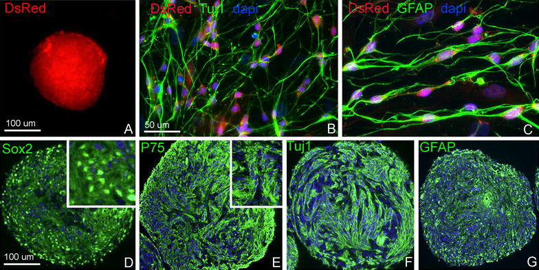
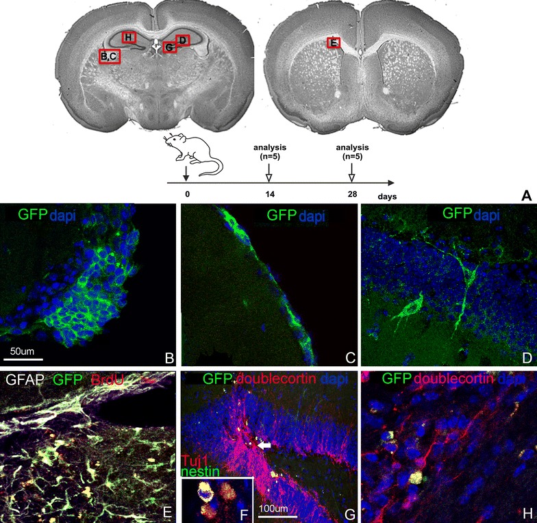
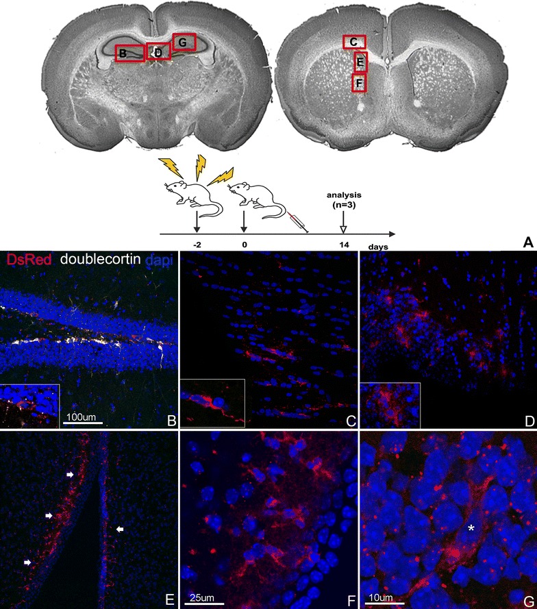
Results: Enteric neuronal stem and progenitor cells, able to differentiate into neuronal and glial lineages, were isolated from the postnatal enteric nervous system and propagated in vitro. Gut-derived neural progenitors, genetically engineered to express fluorescent proteins, were transplanted into the injured brain of adult mice. Using different models of brain injury in combination with either local or systemic cell delivery, we show that transplanted enteric neuronal progenitor cells survive, proliferate, and differentiate into neuronal and glial lineages in vivo. Moreover, transplanted cells migrate extensively along neuronal pathways and appear to modulate the local microenvironment to stimulate endogenous neurogenesis.

Conclusions: Our findings suggest that enteric nervous system derived cells represent a potential source for tissue regeneration in the central nervous system. Further studies are needed to validate these findings and to explore whether autologous gut-derived cell transplantation into the injured brain can result in functional neurologic recovery.


Figures

Fig. 1
Fig. 1
In vitro characterization of multipotent, self-renewing progenitor cells from the postnatal ENS. Enteric neurospheres derived from Actb-DsRed mouse colon express red fluorescent protein (A). Enteric neuronal stem/progenitor cells (ENSCs) within those neurospheres differentiate into neurons (B) and glia (C) that retain DsRed expression. Immunohistochemical characterization shows that cells within the neurospheres express markers of neural progenitors (Sox2, D; P75, E), enteric neurons (Tuj1, F), and glial cells (GFAP, G)
Fig. 2
Fig. 2
Survival and localization of ENSCs following intracerebral delivery. Nestin-GFP+ ENSCs were injected into various brain regions and mice were analyzed at 2 weeks and 4 weeks post transplantation into the brain regions depicted (A). Transplanted cells were found in clusters (B) and monolayers (C) along the ventricular lining, and within the parenchyma of the hippocampus formation (D, FH) and subventricular zone (E). BrdU labeling confirms survival of proliferating ENSCs (E). Differentiation into glial (E) and neuronal (FH) phenotypes is also seen (F represents an inset from the same region as G, with Tuj1 as a marker of mature neurons). Increased numbers of endogenous doublecortin+ neurons within the dentate gyrus are identified in close proximity to GFP+ transplanted cells (G, arrow)
Fig. 3
Fig. 3
Following concussion injury, ENSCs cluster around injured brain parenchyma, migrate to neurogenic niches, and induce focal neurogenesis. Concussion injury was induced over 5 days, ENSCs delivered systemically 3 days later, and brains analyzed after 10 weeks. Brain regions shown in each panel are indicated in (A). ENSCs are identified at the site of brain injury, both at the cortical surface (B, thin arrows) and in the parenchyma (BG). ENSCs appear to be migrating from the injury site toward the ipsilateral dentate gyrus (B, thick arrows). Doublecortin + endogenous neurons are observed in increased numbers in the region of the transplanted cells in the dentate gyrus (EG) and at the cortical surface (H, arrows highlight transplanted ENSCs adjacent to endogenous doublecortin + neurons)
Fig. 4
Fig. 4
Systemically delivered ENSCs home to the brain following whole body radiation. ENSCs were delivered systemically 2 days after whole body radiation and mice analyzed at 14 days. Brain regions shown in each panel are indicated in (A). DsRed + cells are present in the subgranular layer of the dentate gyrus, alongside endogenous doublecortin + (white) neurons (B). Transplanted cells are also found within large white matter tracts bordering the subventricular zone (C), choroid plexus (D), and subependymal layer of the subventricular zone (EG)
Fig. 5
Fig. 5
ENSCs home to the brain and undergo neuronal differentiation after focal brain irradiation. Mice were subjected to focal brain irradiation, transplanted with DsRed + ENSCs via tail vein 2 days later, and analyzed at 28 days. Brain regions shown in each panel are indicated in (A). Transplanted cells are present in the subventricular zone and co-express Hu, consistent with neuronal differentiation (BE, G, H). DsRed + cells are also seen in the dentate gyrus (I), in close proximity to Olig2 + oligodendrocytes (F)

Similar articles

Cited by

References

    1. Mukherjee D, Patil CG. Epidemiology and the global burden of stroke. World Neurosurg. 2011;76(6 Suppl):S85–S90. doi: 10.1016/j.wneu.2011.07.023. - DOI - PubMed
    1. Gustavsson A, Svensson M, Jacobi F, Allgulander C, Alonso J, Beghi E, Dodel R, Ekman M, Faravelli C, Fratiglioni L, et al. Cost of disorders of the brain in Europe 2010. Euro Neuropsychopharmacol J Euro Coll Neuropsychopharmacol. 2011;21(10):718–779. doi: 10.1016/j.euroneuro.2011.08.008. - DOI - PubMed
    1. Wittchen HU, Jacobi F, Rehm J, Gustavsson A, Svensson M, Jonsson B, Olesen J, Allgulander C, Alonso J, Faravelli C, et al. The size and burden of mental disorders and other disorders of the brain in Europe 2010. Euro Neuropsychopharmacol J Euro Coll Neuropsychopharmacol. 2011;21(9):655–679. doi: 10.1016/j.euroneuro.2011.07.018. - DOI - PubMed
    1. Daviglus ML, Bell CC, Berrettini W, Bowen PE, Connolly ES, Jr, Cox NJ, Dunbar-Jacob JM, Granieri EC, Hunt G, McGarry K, et al. National Institutes of Health State-of-the-Science Conference statement: preventing alzheimer disease and cognitive decline. Ann Intern Med. 2010;153(3):176–181. doi: 10.7326/0003-4819-153-3-201008030-00260. - DOI - PubMed
    1. Saraceno B. Neurological disorders and public health challenges. In: Document WHO, editor. Geneva. Switzerland: World Health Organization Press; 2006. pp. 1–218.

MeSH terms

LinkOut - more resources