

Analysis of Glial Distribution in Drosophila Adult Brains
- PMID: 26810782
- PMCID: PMC5563736
- DOI: 10.1007/s12264-016-0014-0
Analysis of Glial Distribution in Drosophila Adult Brains
Abstract
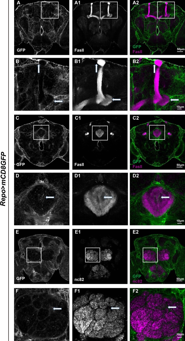
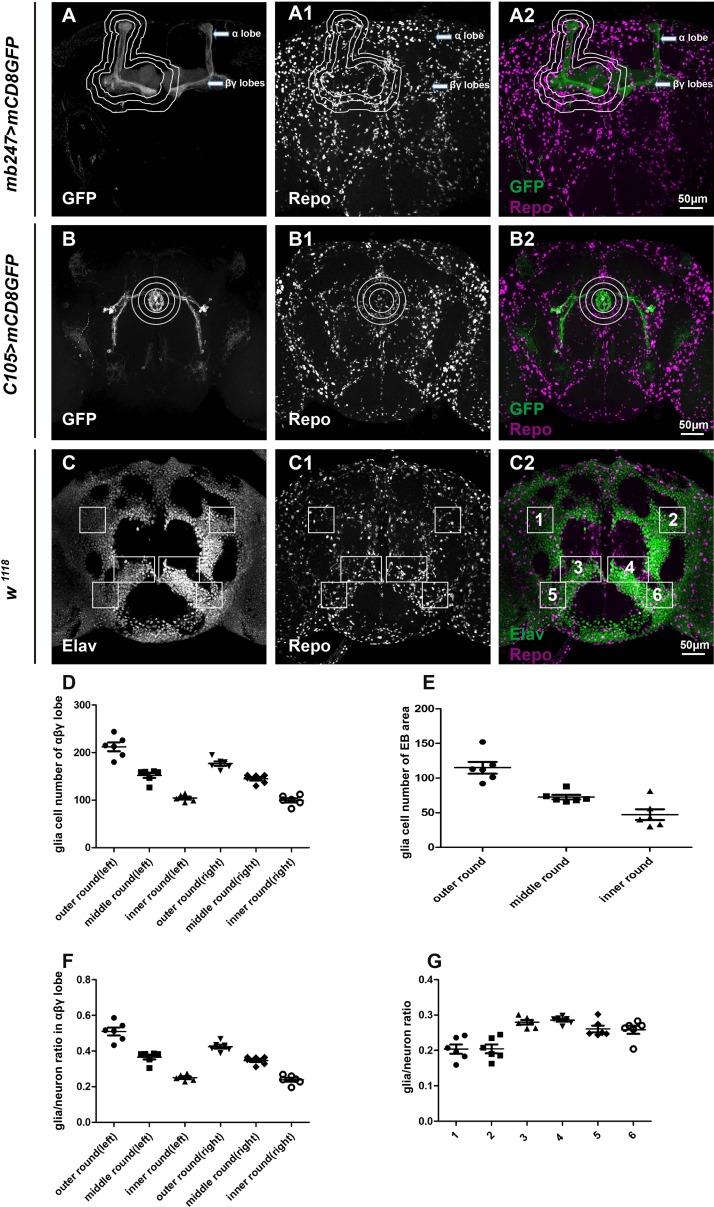
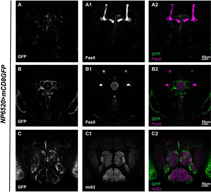
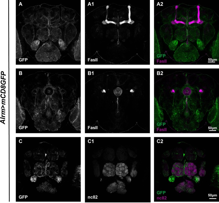
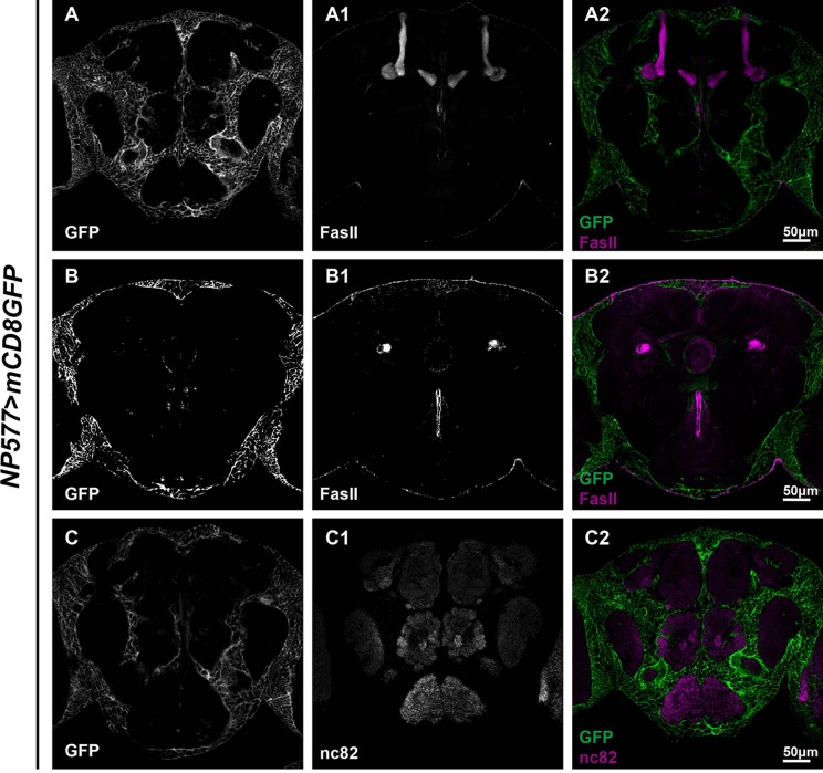
Neurons and glia are the two major cell types in the nervous system and work closely with each other to program neuronal interplay. Traditionally, neurons are thought to be the major cells that actively regulate processes like synapse formation, plasticity, and behavioral output. Glia, on the other hand, serve a more supporting role. To date, accumulating evidence has suggested that glia are active participants in virtually every aspect of neuronal function. Despite this, fundamental features of how glia interact with neurons, and their spatial relationships, remain elusive. Here, we describe the glial cell population in Drosophila adult brains. Glial cells extend and tightly associate their processes with major structures such as the mushroom body (MB), ellipsoid body (EB), and antennal lobe (AL) in the brain. Glial cells are distributed in a more concentrated manner in the MB. Furthermore, subsets of glia exhibit distinctive association patterns around different neuronal structures. Whereas processes extended by astrocyte-like glia and ensheathing glia wrap around the MB and infiltrate into the EB and AL, cortex glia stay where cell bodies of neurons are and remain outside of the synaptic regions structured by EB or AL.
Keywords: Drosophila; Glia; Mushroom body.
Figures
Similar articles
-
The glia of the adult Drosophila nervous system.Glia. 2017 Apr;65(4):606-638. doi: 10.1002/glia.23115. Epub 2017 Jan 30. Glia. 2017. PMID: 28133822 Free PMC article.
-
Organization and postembryonic development of glial cells in the adult central brain of Drosophila.J Neurosci. 2008 Dec 17;28(51):13742-53. doi: 10.1523/JNEUROSCI.4844-08.2008. J Neurosci. 2008. PMID: 19091965 Free PMC article.
-
Astrocyte-like glial cells physiologically regulate olfactory processing through the modification of ORN-PN synaptic strength in Drosophila.Eur J Neurosci. 2014 Sep;40(5):2744-54. doi: 10.1111/ejn.12646. Epub 2014 Jun 26. Eur J Neurosci. 2014. PMID: 24964821
-
Morphological diversity and development of glia in Drosophila.Glia. 2011 Sep;59(9):1237-52. doi: 10.1002/glia.21162. Epub 2011 Mar 24. Glia. 2011. PMID: 21438012 Free PMC article. Review.
-
Neuron-glia interaction in the Drosophila nervous system.Dev Neurobiol. 2021 Jul;81(5):438-452. doi: 10.1002/dneu.22737. Epub 2020 Mar 4. Dev Neurobiol. 2021. PMID: 32096904 Review.
Cited by
-
Gap junction protein Innexin2 modulates the period of free-running rhythms in Drosophila melanogaster.iScience. 2021 Aug 20;24(9):103011. doi: 10.1016/j.isci.2021.103011. eCollection 2021 Sep 24. iScience. 2021. PMID: 34522854 Free PMC article.
-
Experience-dependent glial pruning of synaptic glomeruli during the critical period.Sci Rep. 2024 Apr 20;14(1):9110. doi: 10.1038/s41598-024-59942-3. Sci Rep. 2024. PMID: 38643298 Free PMC article.
-
Glial metabolism versatility regulates mushroom body-driven behavioral output in Drosophila.Learn Mem. 2024 Jun 11;31(5):a053823. doi: 10.1101/lm.053823.123. Print 2024 May. Learn Mem. 2024. PMID: 38862167 Free PMC article. Review.
-
Taurine Transporter dEAAT2 is Required for Auditory Transduction in Drosophila.Neurosci Bull. 2018 Dec;34(6):939-950. doi: 10.1007/s12264-018-0255-1. Epub 2018 Jul 24. Neurosci Bull. 2018. PMID: 30043098 Free PMC article.
-
Alcohol sedation in adult Drosophila is regulated by Cysteine proteinase-1 in cortex glia.Commun Biol. 2019 Jul 3;2:252. doi: 10.1038/s42003-019-0492-5. eCollection 2019. Commun Biol. 2019. PMID: 31286069 Free PMC article.
References
Publication types
MeSH terms
Substances
LinkOut - more resources
Full Text Sources
Other Literature Sources
Molecular Biology Databases
Miscellaneous
