

Time-Lapse Retinal Ganglion Cell Dendritic Field Degeneration Imaged in Organotypic Retinal Explant Culture
- PMID: 26811145
- PMCID: PMC4736988
- DOI: 10.1167/iovs.15-17769
Time-Lapse Retinal Ganglion Cell Dendritic Field Degeneration Imaged in Organotypic Retinal Explant Culture
Abstract
Purpose: To develop an ex vivo organotypic retinal explant culture system suitable for multiple time-point imaging of retinal ganglion cell (RGC) dendritic arbors over a period of 1 week, and capable of detecting dendrite neuroprotection conferred by experimental treatments.
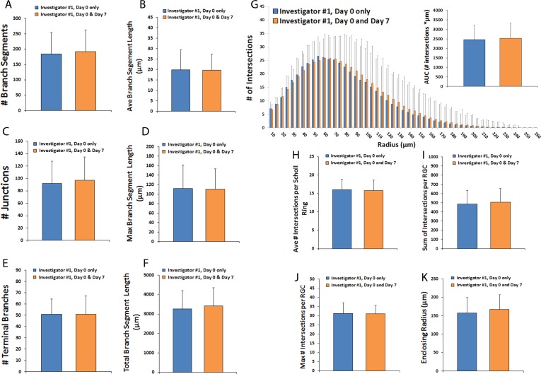
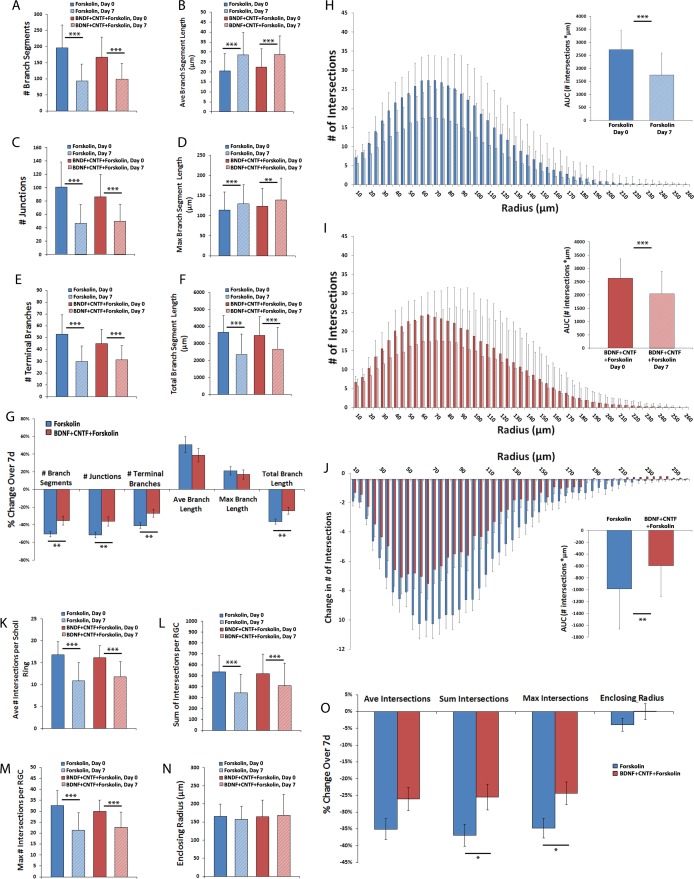
Methods: Thy1-YFP mouse retinas were explanted and maintained in organotypic culture. Retinal ganglion cell dendritic arbors were imaged repeatedly using confocal laser scanning microscopy. Maximal projection z-stacks were traced by two masked investigators and dendritic fields were analyzed for characteristics including branch number, size, and complexity. One group of explants was treated with brain derived neurotrophic factor (BDNF) and ciliary neurotrophic factor (CNTF) added to the culture media. Changes in individual dendritic fields over time were detected using pair-wise comparison testing.
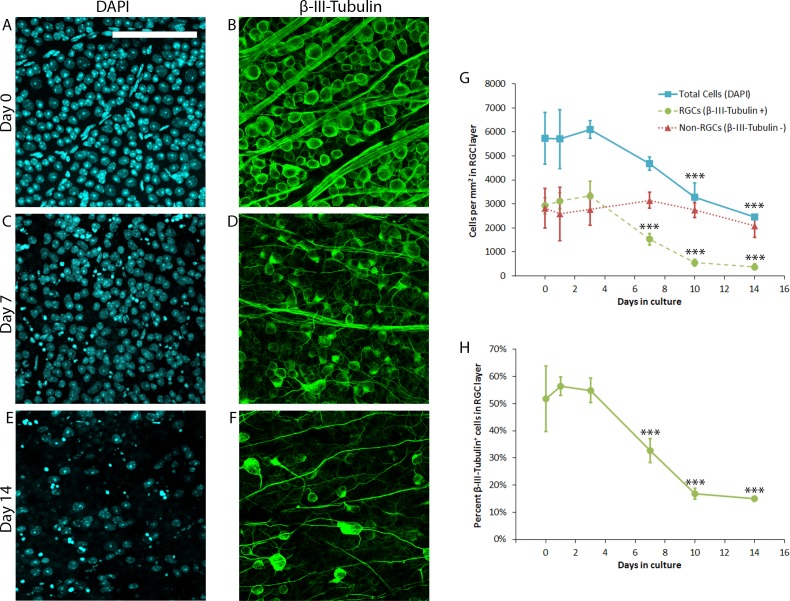
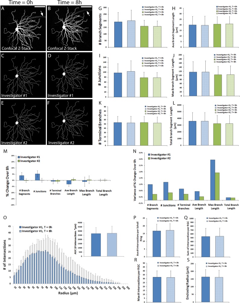
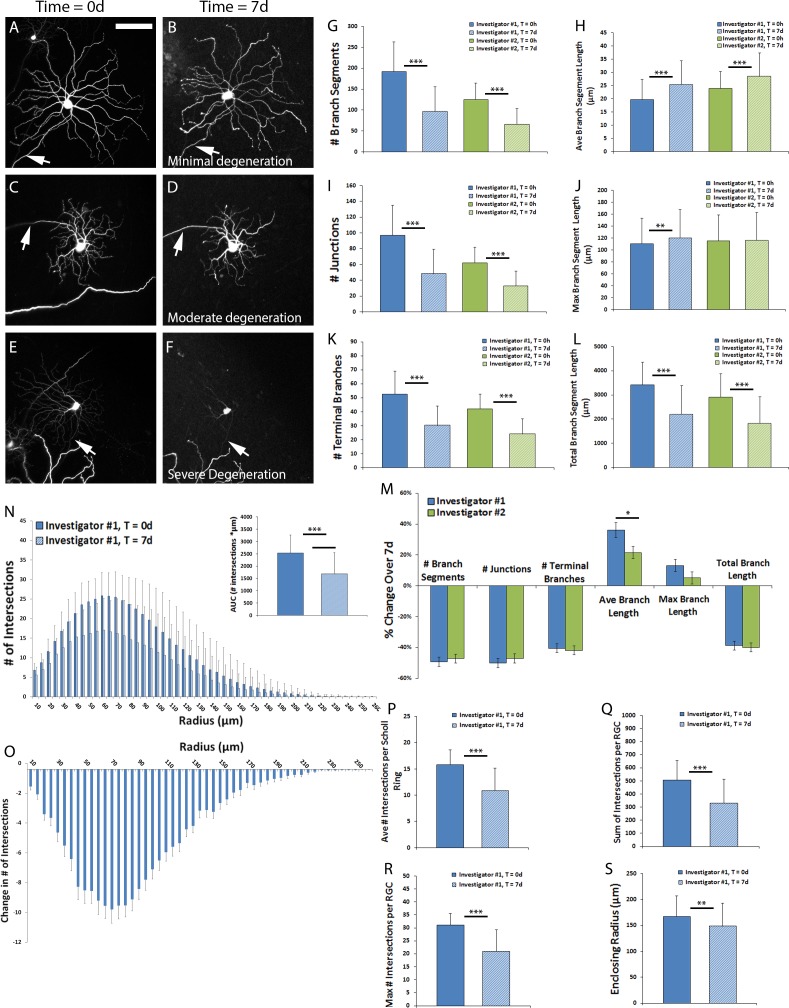
Results: Retinal ganglion cells in mouse retinal explant culture began to degenerate after 3 days with 52.4% surviving at 7 days. Dendritic field parameters showed minimal change over 8 hours in culture. Intra- and interobserver measurements of dendrite characteristics were strongly correlated (Spearman rank correlations consistently > 0.80). Statistically significant (P < 0.001) dendritic tree degeneration was detected following 7 days in culture including: 40% to 50% decreases in number of branch segments, number of junctions, number of terminal branches, and total branch length. Scholl analyses similarly demonstrated a significant decrease in dendritic field complexity. Treatment of explants with BDNF+CNTF significantly attenuated dendritic field degeneration.
Conclusions: Retinal explant culture of Thy1-YFP tissue provides a useful model for time-lapse imaging of RGC dendritic field degeneration over a course of several days, and is capable of detecting neuroprotective amelioration of dendritic pruning within individual RGCs.
Figures
Similar articles
-
Long-term in vivo imaging and measurement of dendritic shrinkage of retinal ganglion cells.Invest Ophthalmol Vis Sci. 2011 Mar 1;52(3):1539-47. doi: 10.1167/iovs.10-6012. Invest Ophthalmol Vis Sci. 2011. PMID: 21245394
-
Tracking dendritic shrinkage of retinal ganglion cells after acute elevation of intraocular pressure.Invest Ophthalmol Vis Sci. 2011 Sep 14;52(10):7205-12. doi: 10.1167/iovs.10-6868. Invest Ophthalmol Vis Sci. 2011. PMID: 21775662
-
Red Light Irradiation In Vivo Upregulates DJ-1 in the Retinal Ganglion Cell Layer and Protects against Axotomy-Related Dendritic Pruning.Int J Mol Sci. 2021 Aug 4;22(16):8380. doi: 10.3390/ijms22168380. Int J Mol Sci. 2021. PMID: 34445085 Free PMC article.
-
Retinal ganglion cell dendrite pathology and synapse loss: Implications for glaucoma.Prog Brain Res. 2015;220:199-216. doi: 10.1016/bs.pbr.2015.04.012. Epub 2015 Jun 30. Prog Brain Res. 2015. PMID: 26497792 Review.
-
Dendritic and synaptic protection: is it enough to save the retinal ganglion cell body and axon?J Neuroophthalmol. 2008 Jun;28(2):144-54. doi: 10.1097/WNO.0b013e318177edf0. J Neuroophthalmol. 2008. PMID: 18562848 Review.
Cited by
-
AAV2-Mediated Transduction of the Mouse Retina After Optic Nerve Injury.Invest Ophthalmol Vis Sci. 2017 Dec 1;58(14):6091-6104. doi: 10.1167/iovs.17-22634. Invest Ophthalmol Vis Sci. 2017. PMID: 29204649 Free PMC article.
-
A mouse ocular explant model that enables the study of living optic nerve head events after acute and chronic intraocular pressure elevation: Focusing on retinal ganglion cell axons and mitochondria.Exp Eye Res. 2017 Jul;160:106-115. doi: 10.1016/j.exer.2017.04.003. Epub 2017 Apr 14. Exp Eye Res. 2017. PMID: 28414059 Free PMC article.
-
Human-Induced Pluripotent Stem Cells Generate Light Responsive Retinal Organoids with Variable and Nutrient-Dependent Efficiency.Stem Cells. 2018 Oct;36(10):1535-1551. doi: 10.1002/stem.2883. Epub 2018 Aug 13. Stem Cells. 2018. PMID: 30004612 Free PMC article.
-
Culture of Adult Transgenic Zebrafish Retinal Explants for Live-cell Imaging by Multiphoton Microscopy.J Vis Exp. 2017 Feb 24;(120):55335. doi: 10.3791/55335. J Vis Exp. 2017. PMID: 28287581 Free PMC article.
-
Role of the Internal Limiting Membrane in Structural Engraftment and Topographic Spacing of Transplanted Human Stem Cell-Derived Retinal Ganglion Cells.Stem Cell Reports. 2021 Jan 12;16(1):149-167. doi: 10.1016/j.stemcr.2020.12.001. Epub 2020 Dec 30. Stem Cell Reports. 2021. PMID: 33382979 Free PMC article.
References
-
- Almasieh M,, Wilson AM,, Morquette B, Cueva Vargas JL, Di Polo A. The molecular basis of retinal ganglion cell death in glaucoma. Prog Retin Eye Res. 2012; 31: 152–181. - PubMed
-
- Tsai JC. Canadian journal of ophthalmology lecture: translational research advances in glaucoma neuroprotection. Can J Ophthalmol. 2013; 48: 141–145. - PubMed
-
- Johnson TV,, Martin KR. Cell transplantation approaches to retinal ganglion cell neuroprotection in glaucoma. Curr Opin Pharmacol. 2013; 13: 78–82. - PubMed
Publication types
MeSH terms
Grants and funding
LinkOut - more resources
Full Text Sources
Other Literature Sources
Miscellaneous
