Review
. 2016 Apr;29(2):155-95.
doi: 10.1007/s10334-015-0521-4. Epub 2016 Jan 25.

A review of heart chamber segmentation for structural and functional analysis using cardiac magnetic resonance imaging

Affiliations
Review

A review of heart chamber segmentation for structural and functional analysis using cardiac magnetic resonance imaging

Peng Peng et al. MAGMA. 2016 Apr.

Abstract

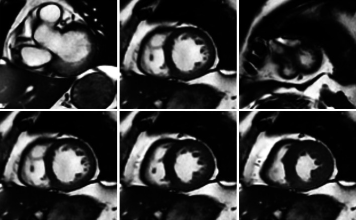
Cardiovascular magnetic resonance (CMR) has become a key imaging modality in clinical cardiology practice due to its unique capabilities for non-invasive imaging of the cardiac chambers and great vessels. A wide range of CMR sequences have been developed to assess various aspects of cardiac structure and function, and significant advances have also been made in terms of imaging quality and acquisition times. A lot of research has been dedicated to the development of global and regional quantitative CMR indices that help the distinction between health and pathology. The goal of this review paper is to discuss the structural and functional CMR indices that have been proposed thus far for clinical assessment of the cardiac chambers. We include indices definitions, the requirements for the calculations, exemplar applications in cardiovascular diseases, and the corresponding normal ranges. Furthermore, we review the most recent state-of-the art techniques for the automatic segmentation of the cardiac boundaries, which are necessary for the calculation of the CMR indices. Finally, we provide a detailed discussion of the existing literature and of the future challenges that need to be addressed to enable a more robust and comprehensive assessment of the cardiac chambers in clinical practice.

Keywords: Cardiac segmentation; Clinical assessment; MRI.


Figures

Fig. 1
Fig. 1
The anatomy of the heart. https://en.wikipedia.org/wiki/Heart
Fig. 2
Fig. 2
Short-axis cine MR images. Top row: slices from base to apex; bottom row mid-cavity slice from diastole to systole, displayed using our automatic cardiac segmentation platform GIMIAS. www.gimias.org
Fig. 3
Fig. 3
LV segmentation in both long-axis and short-axis views [18]
Fig. 4
Fig. 4
Short-axis tagged MRI mid-cavity slices: a tagging produced at end-diastole; bd tag lines deform with myocardial contraction in systole; e, f tag lines deform with myocardial relaxation in diastole; f tag lines fade as the end of a complete cycle is approaching [24]
Fig. 5
Fig. 5
Examples of patients with ischemia acquired in typical late gadolinium enhancement, standard, and high-resolution perfusion MRI. Arrows indicate the inferior scar with thinning of the myocardium [30]
Fig. 6
Fig. 6
End-diastolic (left) and end-systolic (right) myocardial wall thickness measurements on LV SAX mid-cavity slices [48]
Fig. 7
Fig. 7
17-segment model: a recommended myocardial segments and their nomenclatures on a circumferential polar display; b assignment to the territories of the left anterior descending (LAD), right coronary artery (RCA), and the left circumflex coronary artery (LCX). http://www.pharmstresstech.com/stressing/spect.aspx
Fig. 8
Fig. 8
LV endocardium delineation using thresholding: a detected region of interest (ROI); b ROI image; c converted binary image using optimal thresholding [87]
Fig. 9
Fig. 9
Pixel classification by fitting a Gaussian Mixture Model to the histogram of the input image: a the input short-axis image; b 3 Gaussian distributed components representing the air, myocardial muscle, and blood/fat compartment; c the output image with classified pixels in different labels [92]
Fig. 10
Fig. 10
LV epicardium (left) and endocardium (right) tracking: contours propagate through short-axis slices on all phases in a complete cardiac cycle [106]
Fig. 11
Fig. 11
Examples of detected LV myocardial strains visualised in 3D: a ED strain; b ES radial strain; c ES circumferential strain; d ES longitudinal strain [115]
Fig. 12
Fig. 12
A 3D-ASM (SPASM) LV segmentation technique [120] using GIMIAS platform: Step 1 user specifies three landmarks (the aorta, the mitral valve, and the apex) by three clicks on the cine MR volumes; Step 2 the platform automatically generates a model (a triangular surface mesh), which is pre-constructed in training stage, based on the three given landmarks; Step 3 the model fits to the target (feature point detected via fuzzy inference) through propagating the updates from the vertices close to the intersections between the surface and the image planes to distant regions on the earth
Fig. 13
Fig. 13
A framework of ventricular segmentation based on multi-atlas and label fusion technique. Atlases are first registered to the target image. The label at a voxel (red dot) is given by the comparisons between the patch (yellow) on the target image and the patches (colourful boxes) on the warped atlases, weighed by the distance and similarity. Then the fusion of labels from all atlases assigns each voxel a final class. The segmentation result is used to refine the registration process [156]
Fig. 14
Fig. 14
A framework of direct estimation: unsupervised learning searches an efficient image representation way and regression forest trained by using manually segmented data captures the discriminative features [16]
Fig. 15
Fig. 15
An LA blood pool (left) subdivided to Voronoi cells (middle). The narrow junction is the smaller sphere (right) locating between two larger components [163]
Fig. 16
Fig. 16
An evaluation of segmentation accuracy using surface-to-surface (S2S) distance between the segmented result and the manually delineated ground-truth from two different views in 3D [118]
Fig. 17
Fig. 17
The amount of referred publications in each section

References

    1. Alwan A. Global status report on noncommunicable diseases 2010. Geneva: World Health Organization; 2011.
    1. Mendis S, Puska P, Norrving B. Global atlas on cardiovascular disease prevention and control. Geneva: World Health Organization; 2011.
    1. Mathers CD, Loncar D. Projections of global mortality and burden of disease from 2002 to 2030. PLOS Med. 2006;3(11):e442. doi: 10.1371/journal.pmed.0030442. - DOI - PMC - PubMed
    1. Myerson SG, Francis J, Neubauer S. Cardiovascular magnetic resonance. Oxford: OUP; 2013.
    1. Attili AK, Schuster A, Nagel E, Reiber JH, van der Geest RJ. Quantification in cardiac MRI: advances in image acquisition and processing. Int J Cardiovasc Imaging. 2010;26(1):27–40. doi: 10.1007/s10554-009-9571-x. - DOI - PMC - PubMed

Publication types

MeSH terms