. 2016 Mar 25;291(13):6610-24.
doi: 10.1074/jbc.M115.708453. Epub 2016 Jan 25.

PqsBC, a Condensing Enzyme in the Biosynthesis of the Pseudomonas aeruginosa Quinolone Signal: CRYSTAL STRUCTURE, INHIBITION, AND REACTION MECHANISM

Affiliations

PqsBC, a Condensing Enzyme in the Biosynthesis of the Pseudomonas aeruginosa Quinolone Signal: CRYSTAL STRUCTURE, INHIBITION, AND REACTION MECHANISM

Steffen Lorenz Drees et al. J Biol Chem. .

Abstract

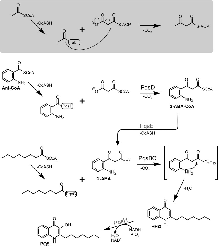
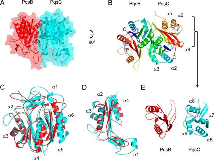
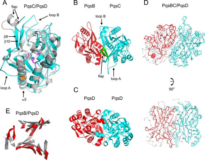
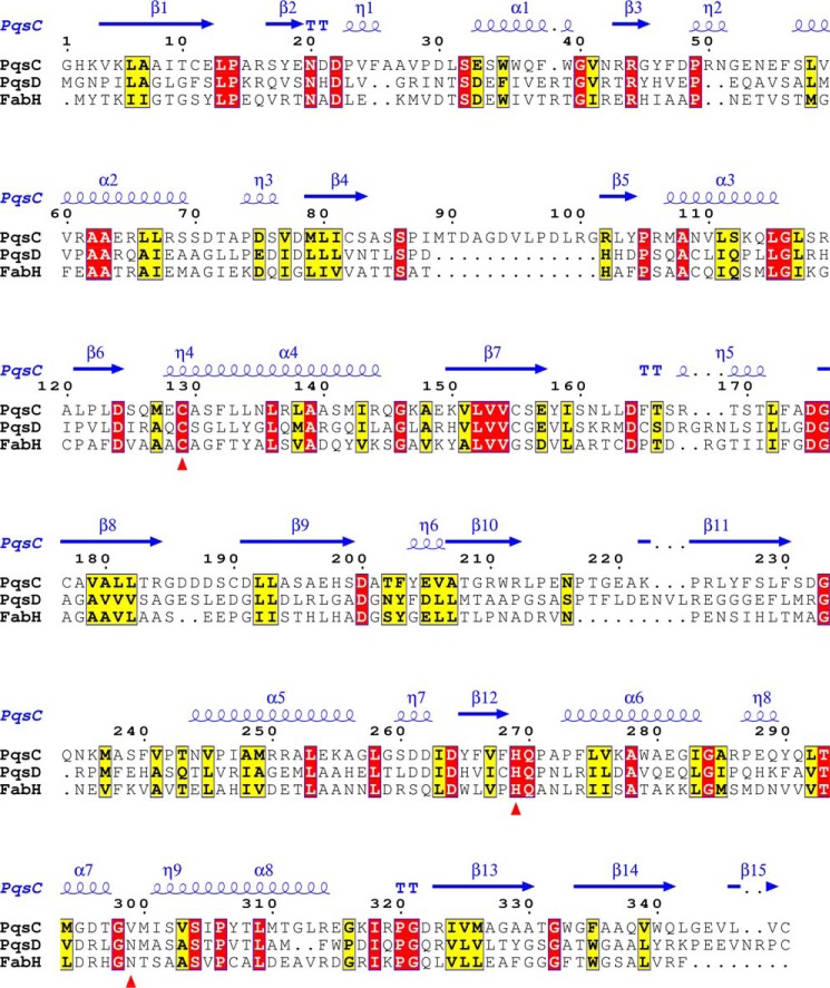
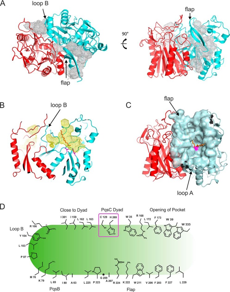
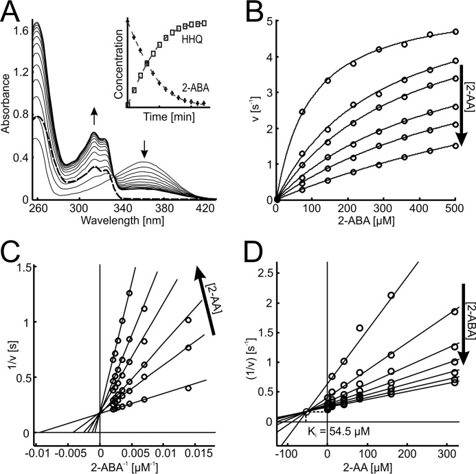
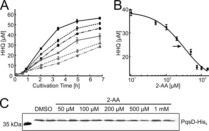
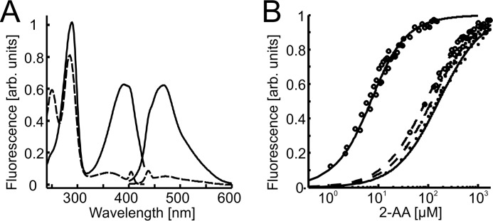
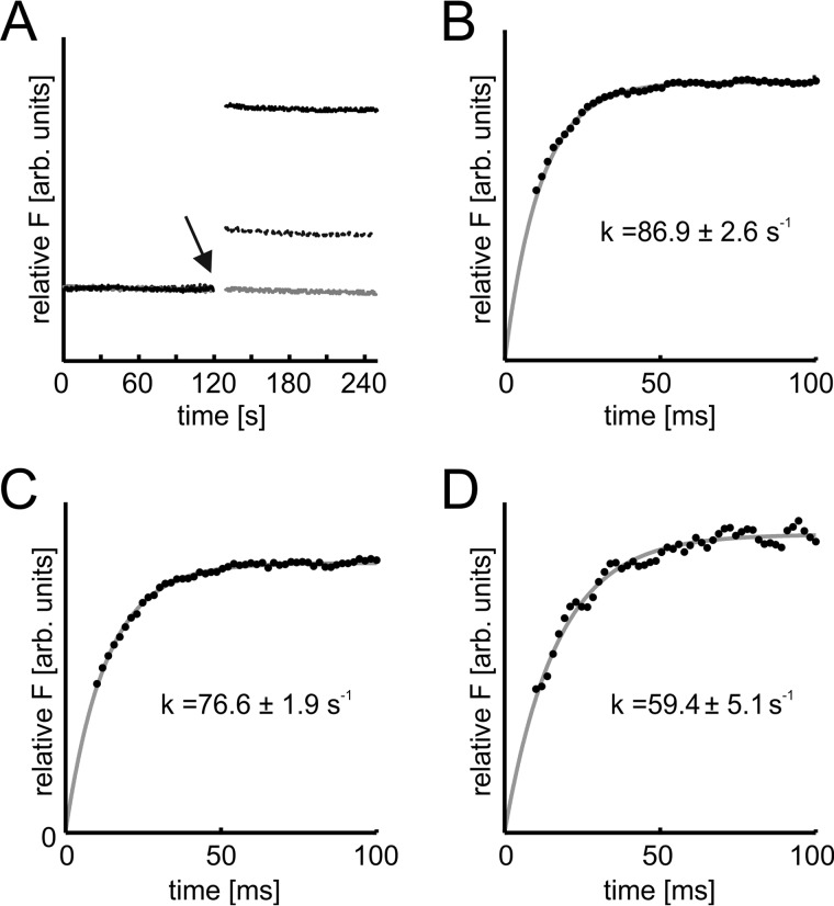
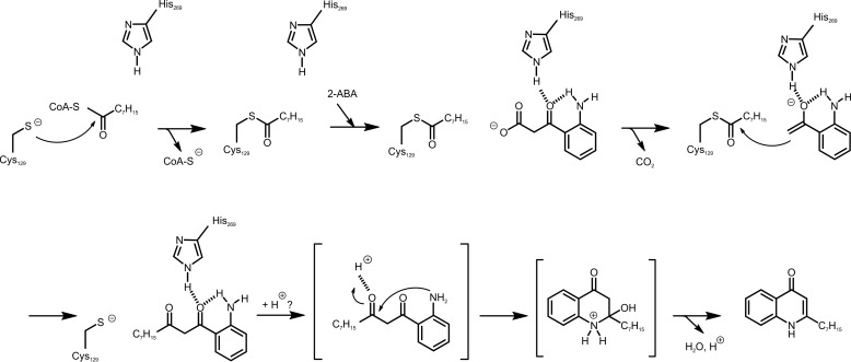
Pseudomonas aeruginosaproduces a number of alkylquinolone-type secondary metabolites best known for their antimicrobial effects and involvement in cell-cell communication. In the alkylquinolone biosynthetic pathway, the β-ketoacyl-(acyl carrier protein) synthase III (FabH)-like enzyme PqsBC catalyzes the condensation of octanoyl-coenzyme A and 2-aminobenzoylacetate (2-ABA) to form the signal molecule 2-heptyl-4(1H)-quinolone. PqsBC, a potential drug target, is unique for its heterodimeric arrangement and an active site different from that of canonical FabH-like enzymes. Considering the sequence dissimilarity between the subunits, a key question was how the two subunits are organized with respect to the active site. In this study, the PqsBC structure was determined to a 2 Å resolution, revealing that PqsB and PqsC have a pseudo-2-fold symmetry that unexpectedly mimics the FabH homodimer. PqsC has an active site composed of Cys-129 and His-269, and the surrounding active site cleft is hydrophobic in character and approximately twice the volume of related FabH enzymes that may be a requirement to accommodate the aromatic substrate 2-ABA. From physiological and kinetic studies, we identified 2-aminoacetophenone as a pathway-inherent competitive inhibitor of PqsBC, whose fluorescence properties could be used forin vitrobinding studies. In a time-resolved setup, we demonstrated that the catalytic histidine is not involved in acyl-enzyme formation, but contributes to an acylation-dependent increase in affinity for the second substrate 2-ABA. Introduction of Asn into the PqsC active site led to significant activity toward the desamino substrate analog benzoylacetate, suggesting that the substrate 2-ABA itself supplies the asparagine-equivalent amino function that assists in catalysis.

Keywords: 2-alkyl-4(1H)-quinolones; FabH; Pseudomonas aeruginosa (P. aeruginosa); Pseudomonas quinolone signal; biosynthesis; condensing enzyme; crystal structure; quorum sensing; secondary metabolism.


Figures

FIGURE 1.
FIGURE 1.
Reactions of FabH (shaded box) and the FabH-like proteins PqsD and PqsBC involved in 2-alkyl-4(1H)-quinolone biosynthesis. The PqsD- and PqsBC-catalyzed reactions proceed in a similar manner to that of the model enzyme FabH. In the initial step, the acyl moiety of an activated carboxylic acid is transferred to a strictly conserved active-site cysteine residue. Subsequently, a β-ketoalkanoic acid (malonyl-ACP in case of FabH, malonyl-CoA or malonyl-ACP in PqsD, and 2-ABA in PqsBC) is decarboxylated, and the resulting reactive enolate intermediate (not shown) attacks the thioester bond of the acyl-enzyme to form the reaction product. Intermediates and products of the alkylquinolone biosynthetic pathway: Ant-CoA, anthraniloyl-coenzyme A; 2-ABA-CoA, 2-aminobenzoylacetyl-CoA; 2-ABA, 2-aminobenzoylacetate; HHQ, 2-heptyl-4(1H)-quinolone; PQS, Pseudomonas quinolone signal.
FIGURE 2.
FIGURE 2.
Schematic diagrams of the PqsBCC129A structure. A, PqsBCC129A heterodimer is shown with PqsC (cyan) and PqsB (red) covered with a transparent molecular surface illustrating the interface. B, PqsB and PqsCC129A structures are colored in a rainbow from the N (blue) to the C terminus (red) with secondary structure elements labeled for PqsCC129A in all panels. C, superposition of PqsB and PqsCC129A. D, N-terminal sub-domains of PqsB(1–154) and PqsC(1–184) superposed with the catalytic dyad residue Ala-129 shown as sticks (purple). E, C-terminal sub-domains of PqsB(155–279) and PqsC(188–348) arranged side by side viewed with the same orientation showing PqsCC129A helix α6 is missing in PqsB. PqsC residue His-269 is shown as sticks (purple).
FIGURE 3.
FIGURE 3.
Stereo figures to illustrate the PqsBCC129A catalytic dyad. A, 2.0 Å electron density map is shown (gray) contoured at 2.0 σ level calculated using the final refined weighted 2FoFc coefficients (REFMAC) and rendered with PyMOL showing PqsC residues Ala-129, Ala-130, His-269, Gln-270, Val-299, and Met-300 as sticks in green. B, PqsBCC129A Ala-129, His-269 catalytic dyad is shown in cyan superposed with the PqsDC112A triad residues (gray).
FIGURE 4.
FIGURE 4.
Comparison of PqsD and PqsBCC129A. A, PqsCC129A structure is shown as a schematic (cyan) superposed onto PqsD (gray). Residues from PqsC shown as sticks in purple are Ala-129, His-269, Val-299, and Pro-242 (orange). PqsD residues Ala-112, His-257, and Asn-287 are shown as sticks (gray). B, schematic diagram of PqsBC (red, cyan) with the flap (PqsC residues 207–227) in green shown in the same orientation as the PqsD homodimer below (C). D, superposition of the PqsBC structure (red, cyan) with the PqsD homodimer (gray) illustrated as Cα-backbone representations with two views shown. E, same as D, but only β-strands for PqsB (red) and PqsD (gray) are shown as schematics.
FIGURE 5.
FIGURE 5.
Alignment of the PqsC amino acid sequence with related enzymes showing the assignments of the PqsCC129A structure at the top (blue). The catalytic residues are marked by red triangles. The sequences shown are PqsD (P. aeruginosa) and FabH (E. coli).
FIGURE 6.
FIGURE 6.
PqsBCC129A active site pocket. A, two views showing the internal pockets calculated using metapocket shown as mesh (gray) together with schematic representations of the PqsBCC129A structure. B, PqsBC structure is shown with the view clipped such that the PqsC flap is removed to show the relationship between the PqsC catalytic dyad (purple) and the PqsC cavity (yellow mesh). For perspective, a decyl formate molecule is shown as sticks (orange) derived from a superposition of the Mtb FabH-decyl formate complex structure (PDB code 2QNZ). C, transparent surface representation of the PqsC active site pocket is show (light cyan) with residue Ala-129 in purple. For perspective, the decyl formate molecules showing the orientation of the proposed entry and exit portals from the FabH-decyl formate complex structure are shown (orange). D, graphical representation of the PqsBC active site cleft. Residues conserved with CoA-binding sites of FabH/PqsD enzymes are Trp-35 and Arg-168.
FIGURE 7.
FIGURE 7.
Steady-state kinetics and competitive inhibition of PqsBC by 2-AA. A, conversion of 2-ABA (λmax at 360 nm) and octanoyl-CoA (absorbing at 260 nm) to HHQ (λmax at 313 nm). 20 nm PqsBC was mixed with 50 μm of each substrate. Spectra were measured every 60 s with a bandwidth of 1 nm. HHQ formation could be observed at 313 nm with a differential extinction coefficient of 6,520 m−1 cm−1. The remaining absorption above 340 nm after full conversion of the substrates (bold trace) is due to scattering and can be eliminated by diluting to 50% in 2-propanol/HCl (dashed trace). Inset, conversion of 10 μm substrates, monitored at 313 nm (HHQ) and 360 nm (2-ABA) with the determined extinction coefficients. Data interval is 1 min. B–D, inhibition of PqsBC activity by 2-AA. B, nonlinear fits of measured initial rates. The Lineweaver-Burk plot (C) shows the characteristic pattern of competitive inhibition. Using the Dixon plot (D), the inhibitor constant Ki of the competitive reaction could be determined graphically. Experiments were performed with 50 μm octanoyl-CoA and 20 nm PqsBC in HEPES buffer, pH 8.2, at 25 °C; arrows indicate ascending concentrations.
FIGURE 8.
FIGURE 8.
In vivo inhibition of HHQ biosynthesis by 2-AA. Cultures of P. putida KT2440 [pBBR::pqsABCD-his] were grown in the presence of 1 mm anthranilic acid (as precursor for HHQ synthesis) and various concentrations of 2-AA. 2-AA does not affect growth of P. putida KT2440, and it is not utilized as a carbon source. A, HHQ contents of culture samples, as determined by HPLC. Black solid line, DMSO control; black dashed line, 50 μm; black dash-dotted line, 100 μm; gray solid line, 200 μm; gray dashed line, 500 μm; gray dash-dotted line, 1 mm 2-AA. Errors reflect standard errors (S.E.) of three biological replicates. B, dose-response plot, using cultures sampled 3.5 h after inoculation. The arrow indicates the calculated EC50 of 319 ± 38 μm. C, immunodetection of PqsD-His6 in Western blots of cell extract supernatants confirms synthesis of the Pqs proteins. Cell extracts were from cultures (independent duplicates) harvested after 7 h of incubation. Each lane contained 50 μg of total protein.
FIGURE 9.
FIGURE 9.
Equilibrium binding of 2-AA to PqsBC proteins in apo- and octanoylated forms. A, fluorescence excitation and emission spectra of 2-AA (1 μm) in unbound (dashed line) and protein-bound (20 μm PqsBC, continuous line) form. B, titration data and curve fittings of 5 μm PqsBC (continuous lines) or PqsBCH269A variant (dashed lines) with 2-AA in the presence (circles) or absence (dots) of 10 μm octanoyl-CoA. Data suggest an increase of binding affinity to 2-AA of the acyl-protein in the wild-type but not in the H269A variant (numerical data provided in Table 2).
FIGURE 10.
FIGURE 10.
Role of His-269 in octanoylation of PqsBC, monitored by transient kinetics. A, time-dependent measurement of 2-AA fluorescence (λEx = 400 nm, λEm = 460 nm, 0.5-s time resolution) in complexes with PqsBC proteins. Octanoylation (addition of 10-fold molar excess of octanoyl-CoA indicated by arrow) shifts the binding equilibrium of 2-AA and PqsBC (solid black line). The effect was reduced in PqsBCH269A (dashed black line), and addition of octanoyl-CoA to PqsBCC129A did not change 2-AA fluorescence (gray line). Enzyme and 2-AA concentrations were varied between 1–20 and 1–100 μm respectively. B–D, octanoylation of PqsBC proteins, analyzed with stopped-flow fluorimetry. B, PqsBC octanoylation probed with 2-AA. C, PqsBCH269A, probed with 2-AA. D, PqsBCH269A, probed with 2-ABA. Assays were conducted at 6 °C, detector response time was 10 ms; enzyme concentration was varied to achieve sufficient signal to noise ratio. Rate constants were calculated assuming a first order reaction characteristic. Denoted errors are standard errors of cross-validation.
FIGURE 11.
FIGURE 11.
Proposed catalytic mechanism of PqsBC toward the formation of the 1-(2-aminophenyl)decane-1,3-dione intermediate and its reaction to HHQ. For details, see text.

References

    1. Lyczak J. B., Cannon C. L., and Pier G. B. (2000) Establishment of Pseudomonas aeruginosa infection: lessons from a versatile opportunist. Microbes Infect. 2, 1051–1060 - PubMed
    1. Williams P. (2007) Quorum sensing, communication and cross-kingdom signalling in the bacterial world. Microbiology 153, 3923–3938 - PubMed
    1. Storz M. P., Maurer C. K., Zimmer C., Wagner N., Brengel C., de Jong J. C., Lucas S., Müsken M., Häussler S., Steinbach A., and Hartmann R. W. (2012) Validation of PqsD as an anti-biofilm target in Pseudomonas aeruginosa by development of small-molecule inhibitors. J. Am. Chem. Soc. 134, 16143–16146 - PubMed
    1. Starkey M., Lepine F., Maura D., Bandyopadhaya A., Lesic B., He J., Kitao T., Righi V., Milot S., Tzika A., and Rahme L. (2014) Identification of anti-virulence compounds that disrupt quorum-sensing regulated acute and persistent pathogenicity. PLoS Pathog. 10, e1004321. - PMC - PubMed
    1. Lu C., Maurer C. K., Kirsch B., Steinbach A., and Hartmann R. W. (2014) Overcoming the unexpected functional inversion of a PqsR antagonist in Pseudomonas aeruginosa: an in vivo potent antivirulence agent targeting pqs quorum sensing. Angew. Chem. Int. Ed. Engl. 53, 1109–1112 - PubMed

Publication types

MeSH terms

LinkOut - more resources