. 2016 Mar 1;143(5):764-73.
doi: 10.1242/dev.126656. Epub 2016 Jan 25.

BMP signaling and cellular dynamics during regeneration of airway epithelium from basal progenitors

Affiliations

BMP signaling and cellular dynamics during regeneration of airway epithelium from basal progenitors

Tomomi Tadokoro et al. Development. .

Abstract

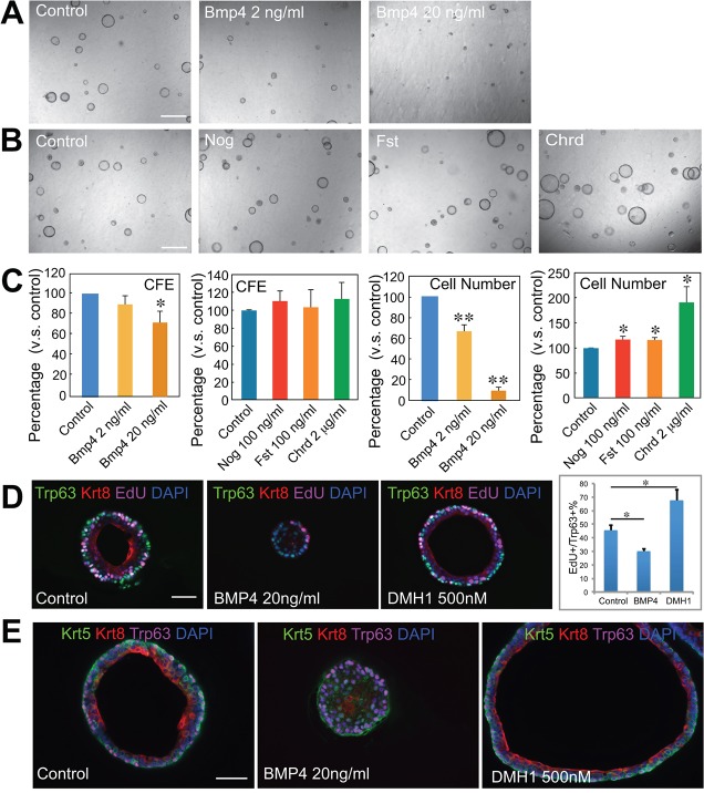
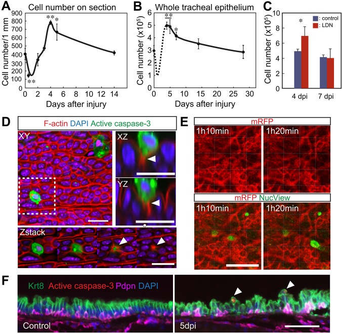
The pseudostratified epithelium of the lung contains ciliated and secretory luminal cells and basal stem/progenitor cells. To identify signals controlling basal cell behavior we screened factors that alter their self-renewal and differentiation in a clonal organoid (tracheosphere) assay. This revealed that inhibitors of the canonical BMP signaling pathway promote proliferation but do not affect lineage choice, whereas exogenous Bmp4 inhibits proliferation and differentiation. We therefore followed changes in BMP pathway components in vivo in the mouse trachea during epithelial regeneration from basal cells after injury. The findings suggest that BMP signaling normally constrains proliferation at steady state and this brake is released transiently during repair by the upregulation of endogenous BMP antagonists. Early in repair, the packing of epithelial cells along the basal lamina increases, but density is later restored by active extrusion of apoptotic cells. Systemic administration of the BMP antagonist LDN-193189 during repair initially increases epithelial cell number but, following the shedding phase, normal density is restored. Taken together, these results reveal crucial roles for both BMP signaling and cell shedding in homeostasis of the respiratory epithelium.

Keywords: Airway epithelium; Apoptosis; BMP signaling; Basal cells; Cell shedding; Homeostasis; Regeneration; SO2 injury.


Conflict of interest statement

Competing interests

The authors declare no competing or financial interests.

Figures

Fig. 1.
Fig. 1.
BMP inhibitors promote cell proliferation of tracheal basal cells. (A) Assay schematic. Ngfr+ basal cells (BCs) were cultured with test compounds in 50% Matrigel in 24-well inserts. The numbers of spheres and cells were quantified at day 9. To the right is a representative bright-field image of spheres at day 9. (B) The effect of potentiators/inhibitors of different signaling pathways in the assay. Bars show cell number as a percentage of the control. Data are the mean of duplicates. The red arrow indicates highest response. (C) Schematic of BMP signaling and inhibitors. LDN-193189 inhibits both BMP and VEGF signaling, whereas DMH1 and DMH2 show little or no effect on VEGF signaling. (D) Bright-field images of spheres in cultures with BMP inhibitors LDN-193189, DMH1 and DMH2. (E) The effect of BMP inhibitors on colony forming efficiency (CFE) and cell number. *P<0.01, **P<0.003 versus control (n=3). Scale bar: 500 µm.
Fig. 2.
Fig. 2.
BMP signaling regulates proliferation of tracheal BCs. (A,B) Bright-field images of spheres treated for 9 days with (A) different concentrations of Bmp4 and (B) the BMP antagonists Nog, Fst and Chrd. (C) Effect of Bmp4 and BMP antagonists on CFE (left) and cell number (right). *P<0.01, **P<0.001 versus control (n=3). (D) Sections of spheres cultured for 7 days under different conditions and exposed to EdU for 2 h before harvest stained with antibodies to Trp63, Krt8 and EdU. The bar chart shows the percentage of Trp63+ cells that are also EdU+. *P<0.05. (E) Sections of spheres cultured for 9 days under different conditions stained with antibodies to Krt5, Krt8 and Trp63. Scale bars: 500 µm in A,B; 50 µm in D,E.
Fig. 3.
Fig. 3.
Dynamic changes in BMP signaling during tissue regeneration. (A) Schematic of repair of tracheal epithelium after SO2 injury. Luminal cells are sloughed off during the first 6-12 h after SO2 exposure (hpi) and BCs spread to cover the denuded area by 24 hpi. BCs proliferate and generate Krt8+ suprabasal descendants that accumulate and become multilayered during the first 6 days. Some differentiated ciliated and secretory cells are first detected around day 3 and regeneration of the epithelium is complete by 2 weeks. Evidence is presented here for cell shedding to restore homeostasis. (B) Phospho-Smad1/5/8 (red) levels in DAPI-stained nuclei of epithelium and mesenchyme during repair after SO2 inhalation. Phospho-Smad1/5/8 is seen in both Trp63+ BCs (green) and luminal cells. Note that not all cells are positive for phospho-Smad1/5/8. Bottom right panels show phospho-Smad1/5/8 in the intercartilage mesenchyme of uninjured tracheas. The cartilage is outlined (dashed line). Some phospho-Smad1/5/8+ cells are also positive for Pdgfra or Bmp4 (green). Scale bars: 50 µm. (C) Quantification of western blot analysis (Fig. S3) of phospho-Smad1/5/8, phospho-Jun and phospho-p38 in the total trachea before (control) and 48 h after (left) injury and phospho-Smad1/5/8 in the epithelium versus mesenchyme before and 48 h after injury (right). Values are the mean of triplicate (left) or duplicate (right) samples. *P<0.05.
Fig. 4.
Fig. 4.
Expression of Bmp-related genes during tissue regeneration. (A) Quantitative RT-PCR analysis of transcripts for genes encoding some BMP ligands, receptors and antagonists in total trachea before and after injury. (B) Bmp4-nCFP expression (red) in trachea before and after injury. Arrowhead indicates weak expression of Bmp4 in the epithelium above Trp63+ BCs (green). (C) After injury, Fst transcripts (red) are detected by in situ hybridization in both the Krt5+ BCs (green) and mesenchyme. **P<0.01 versus uninjured (n=3). Scale bars: 20 μm.
Fig. 5.
Fig. 5.
Inhibition of BMP signaling promotes clonal expansion of BCs. (A) Schematic of clonal analysis of BCs in vivo. Krt5+ BCs were labeled clonally with a low dose of tamoxifen (2.5 µg/g body weight). One week later, mice were given 5% DMSO or 3 mg/kg body weight LDN-193189 by i.p. injection and exposed to SO2 for 4 h. Mice were then treated with drug every 24 h and tracheas harvested at 3 dpi. (B) Schematic of clonal expansion of BCs after injury. Individual BCs were labeled at steady state (red) and clones expanded after injury. Both single cells and clusters were considered to be ‘clones'. (C) (Top) Clone size (cell number/clone) at 3 dpi with and without LDN-193189 treatment. Red bars show the average number of cells/clone: 3.3 for control and 5.8 for LDN-193189-treated mice, respectively. Data are from three mice. *P=5.478×10⁻¹⁶ by Mann–Whitney–Wilcoxon test. (Bottom) Whole-mount image of tracheal epithelium from Krt5-CreER;Rosa-Tomato mouse that had received a low dose of tamoxifen, showing typical clone distribution and size 3 days after SO2 injury. Inset shows higher magnification of the clone in the boxed area. Scale bar: 200 μm at low magnification and 50 μm at high magnification.
Fig. 6.
Fig. 6.
Regulation of cell number in tracheal epithelium by cell extrusion during repair. (A) Number of epithelial cells per mm along basal lamina after injury (n=3 mice). (B) Total epithelial cell number in a single trachea (n=3 mice). The number at day 1 is estimated from the data in A. (C) Total tracheal cell number at 4 dpi and 7 dpi with or without systemic LDN-193189 treatment (n=3 tracheas for control and n=5 tracheas for LDN-193189 treated). *P<0.05, **P<0.01 versus uninjured (n=3). (D) Confocal images of whole tracheal epithelium at 6 dpi after immunohistochemistry showing apoptotic cells (cleaved caspase 3+, green) and F-actin (red). The lower panels show images at different levels of the region boxed in the upper left panel. Upper right panels are enlarged images of an apoptotic cell being extruded. Arrowheads indicate the actin ring in neighboring cells. (E) Snapshots from live cell imaging of tracheal epithelium of a Rosa-mT/mG mouse (red marks epithelial cell membranes) exposed to the caspase 3 substrate Nucview (green). (F) Sections of trachea before injury and at 5 dpi stained with antibodies to Krt8 (luminal cells), Pdpn (BCs) and active caspase 3. The ratio of Krt8+ to Pdpn+ cells is 1.50±0.34 in the control (total cells counted=642) compared with 2.3±0.38 at 5 dpi (total cells counted=735). Arrowheads mark the site of cell extrusion. Scale bars: 20 µm in D; 30 µm in E; 50 μm in F.
Fig. 7.
Fig. 7.
Proposed role of BMP signaling during repair of the tracheal epithelium. At steady state, Bmp4, expressed mainly in the mesenchyme, maintains a low rate of cell proliferation in at least a subset of the BC population. After injury, the BMP antagonist Fst is transiently upregulated both in surviving epithelium and mesenchyme. This, in turn, leads to enhanced epithelial proliferation and differentiation into luminal cells. Cell crowding leads to extrusion and shedding of apoptotic cells and ectopic BMP inhibitors do not increase the final cell density.

Similar articles

Cited by

References

    1. Akiyama I., Yoshino O., Osuga Y., Izumi G., Urata Y., Hirota Y., Hirata T., Harada M., Koga K., Ogawa K. et al. (2013). Follistatin is induced by IL-1beta and TNF-alpha in stromal cells from endometrioma. Reprod. Sci. 20, 675-679. 10.1177/1933719112463253 - DOI - PubMed
    1. Boergermann J. H., Kopf J., Yu P. B. and Knaus P. (2010). Dorsomorphin and LDN-193189 inhibit BMP-mediated Smad, p38 and Akt signalling in C2C12 cells. Int. J. Biochem. Cell Biol. 42, 1802-1807. 10.1016/j.biocel.2010.07.018 - DOI - PMC - PubMed
    1. Borthwick D. W., Shahbazian M., Krantz Q. T., Dorin J. R. and Randell S. H. (2001). Evidence for stem-cell niches in the tracheal epithelium. Am. J. Respir. Cell Mol. Biol. 24, 662-670. 10.1165/ajrcmb.24.6.4217 - DOI - PubMed
    1. Brazil D. P., Church R. H., Surae S., Godson C. and Martin F. (2015). BMP signalling: agony and antagony in the family. Trends Cell Biol. 25, 249-264. 10.1016/j.tcb.2014.12.004 - DOI - PubMed
    1. Chen C. -C., Wang L., Plikus M. V., Jiang T. X., Murray P. J., Ramos R., Guerrero-Juarez C. F., Hughes M. W., Lee O. K., Shi S. et al. (2015). Organ-level quorum sensing directs regeneration in hair stem cell populations. Cell 161, 277-290. 10.1016/j.cell.2015.02.016 - DOI - PMC - PubMed

Publication types

MeSH terms