. 2016 Mar 1;143(5):774-9.
doi: 10.1242/dev.126383. Epub 2016 Jan 25.

MEF2C regulates outflow tract alignment and transcriptional control of Tdgf1

Affiliations

MEF2C regulates outflow tract alignment and transcriptional control of Tdgf1

Ralston M Barnes et al. Development. .

Abstract

Congenital heart defects are the most common birth defects in humans, and those that affect the proper alignment of the outflow tracts and septation of the ventricles are a highly significant cause of morbidity and mortality in infants. A late differentiating population of cardiac progenitors, referred to as the anterior second heart field (AHF), gives rise to the outflow tract and the majority of the right ventricle and provides an embryological context for understanding cardiac outflow tract alignment and membranous ventricular septal defects. However, the transcriptional pathways controlling AHF development and their roles in congenital heart defects remain incompletely elucidated. Here, we inactivated the gene encoding the transcription factor MEF2C in the AHF in mice. Loss of Mef2c function in the AHF results in a spectrum of outflow tract alignment defects ranging from overriding aorta to double-outlet right ventricle and dextro-transposition of the great arteries. We identify Tdgf1, which encodes a Nodal co-receptor (also known as Cripto), as a direct transcriptional target of MEF2C in the outflow tract via an AHF-restricted Tdgf1 enhancer. Importantly, both the MEF2C and TDGF1 genes are associated with congenital heart defects in humans. Thus, these studies establish a direct transcriptional pathway between the core cardiac transcription factor MEF2C and the human congenital heart disease gene TDGF1. Moreover, we found a range of outflow tract alignment defects resulting from a single genetic lesion, supporting the idea that AHF-derived outflow tract alignment defects may constitute an embryological spectrum rather than distinct anomalies.

Keywords: Cripto; Enhancer; Heart development; MEF2; Mouse; Tdgf1.


Conflict of interest statement

Competing interests

The authors declare no competing or financial interests.

Figures

Fig. 1.
Fig. 1.
Mef2c function in the AHF is required for proper OFT alignment. (Aa-d) Normal alignment of the aorta (Ao) and pulmonary artery (PA) in control neonatal mice. RV, right ventricle; LV, left ventricle. (B-D) Subsets of Mef2cAHF-KO neonatal mice displayed a tetralogy of Fallot-like phenotype, including hypoplastic pulmonary artery, ventricular septal defect (VSD, arrowheads), overriding aorta, and hypertrophic right ventricle (Ba-d); dextro-transposition of the great arteries (d-TGA) with VSD (Ca-d); or double-outlet right ventricle (DORV) with or without d-TGA (Da-d). Note that all Mef2cAHF-KO mice displayed thickened and club-shaped semilunar valves, as shown in Bd-Dd.
Fig. 2.
Fig. 2.
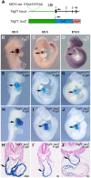
Tdgf1 is expressed in the AHF and the activity of a cardiac-specific enhancer mimics endogenous expression. (A) The murine Tdgf1 locus and Tdgf1::lacZ reporter transgene. The canonical MEF2 binding site in the enhancer is indicated. Exons are numbered. (B-D) Whole-mount in situ hybridization for Tdgf1 in wild type (wt) at E8.5 (B), E9.5 (C), E10.5 (D). (E-G) X-gal staining of Tdgf1lacZ/+ embryos also shows endogenous expression of Tdgf1 from the knock-in allele at E8.5 (E), E9.5 (F), E10.5 (G). (H-J) X-gal-stained embryos of a stable Tdgf1::lacZ transgenic line at E8.5 (H,H′), E9.5 (I,I′), E10.5 (J,J′). (H′-J′) Transverse sections of X-gal-stained Tdgf1::lacZ transgenic embryos. Arrows indicate the OFT in all panels. Scale bars: 100 µm.
Fig. 3.
Fig. 3.
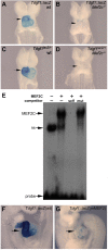
MEF2C is required for Tdgf1 enhancer activity and expression in the heart. (A-D) Tdgf1::lacZ (A,B) and Tdgf1lacZ/+ (C,D) transgenic embryos crossed onto wild-type (wt) (A,C) or Mef2c-null (B,D) backgrounds, collected at E8.5 and X-gal stained. Note the loss of X-gal staining in the absence of Mef2c function (B,D). (E) EMSA of a double-stranded radiolabeled oligonucleotide probe encompassing the Tdgf1 MEF2 site by recombinant MEF2C (lane 2). MEF2C binding was efficiently competed by ∼30-fold excess of unlabeled self probe (lane 3), but was not competed by ∼30-fold excess of a mutant version of the probe (lane 4). Unprogrammed lysate (lacking MEF2C) is shown in lane 1. Bound MEF2C, a non-specific complex (ns) and free probe bands are indicated. (F,G) Representative X-gal-stained E9.5 embryos harboring a wild-type (wt) Tdgf1::lacZ transgene (F) and a Tdgf1::lacZ transgene with a disrupted MEF2 site (mMEF2) (G). 8/13 Tdgf1::lacZ(wt) lines/embryos showed a strong pattern of X-gal staining, such as in the embryo shown in F. By contrast, 2/7 independent Tdgf1::lacZ(mMEF2) founder embryos showed weak X-gal staining, such as shown in G, and 5/7 Tdgf1::lacZ(mMEF2) founder embryos showed no detectable X-gal staining. Arrows (A-D,F,G) mark the OFT.

References

    1. Ai D., Liu W., Ma L., Dong F., Lu M.-F., Wang D., Verzi M. P., Cai C., Gage P. J., Evans S. et al. (2006). Pitx2 regulates cardiac left–right asymmetry by patterning second cardiac lineage-derived myocardium. Dev. Biol. 296, 437-449. 10.1016/j.ydbio.2006.06.009 - DOI - PMC - PubMed
    1. Anderson R. H., Wilkinson J. L., Arnold R., Becker A. E. and Lubkiewicz K. (1974). Morphogenesis of bulboventricular malformations. II. Observations on malformed hearts. Br. Heart J. 36, 948-970. 10.1136/hrt.36.10.948 - DOI - PMC - PubMed
    1. Anderson R. H., Spicer D. E., Giroud J. M. and Mohun T. J. (2013). Tetralogy of Fallot: nosological, morphological, and morphogenetic considerations. Cardiol. Young 23, 858-866. 10.1017/S1047951113001686 - DOI - PubMed
    1. Anderson C. M., Hu J., Barnes R. M., Heidt A. B., Cornelissen I. and Black B. L. (2015). Myocyte enhancer factor 2C function in skeletal muscle is required for normal growth and glucose metabolism in mice. Skelet. Muscle 5, 7 10.1186/s13395-015-0031-0 - DOI - PMC - PubMed
    1. Behrens A. N., Ren Y., Ferdous A., Garry D. J. and Martin C. M. (2012). Nkx2-5 regulates Tdgf1 (Cripto) early during cardiac development. J. Clin. Exp. Cardiol. Suppl. 11, 1-4. - PMC - PubMed

Publication types

MeSH terms