. 2016 Mar 1;143(5):754-63.
doi: 10.1242/dev.129627. Epub 2016 Jan 25.

Chinmo is sufficient to induce male fate in somatic cells of the adult Drosophila ovary

Affiliations

Chinmo is sufficient to induce male fate in somatic cells of the adult Drosophila ovary

Qing Ma et al. Development. .

Abstract

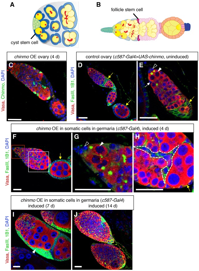
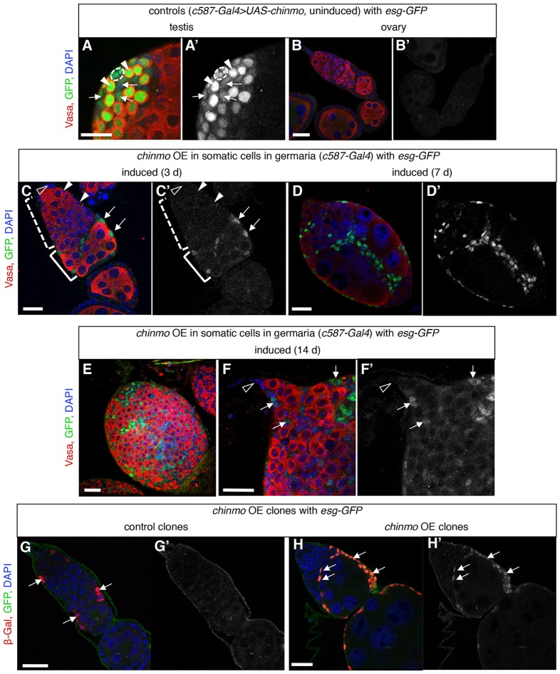
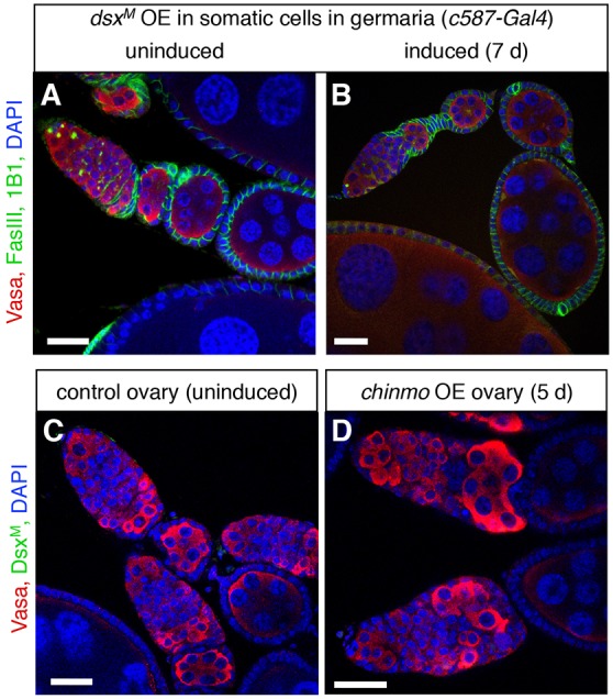
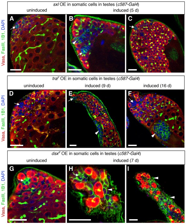
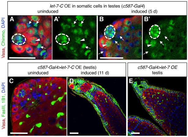
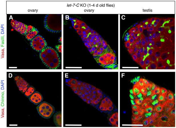
Sexual identity is continuously maintained in specific differentiated cell types long after sex determination occurs during development. In the adult Drosophila testis, the putative transcription factor Chronologically inappropriate morphogenesis (Chinmo) acts with the canonical male sex determinant DoublesexM (Dsx(M)) to maintain the male identity of somatic cyst stem cells and their progeny. Here we find that ectopic expression of chinmo is sufficient to induce a male identity in adult ovarian somatic cells, but it acts through a Dsx(M)-independent mechanism. Conversely, the feminization of the testis somatic stem cell lineage caused by loss of chinmo is enhanced by expression of the canonical female sex determinant Dsx(F), indicating that chinmo acts in parallel with the canonical sex determination pathway to maintain the male identity of testis somatic cells. Consistent with this finding, ectopic expression of female sex determinants in the adult testis disrupts tissue morphology. The miRNA let-7 downregulates chinmo in many contexts, and ectopic expression of let-7 in the adult testis is sufficient to recapitulate the chinmo loss-of-function phenotype, but we find no apparent phenotypes upon removal of let-7 in the adult ovary or testis. Our finding that chinmo is necessary and sufficient to promote a male identity in adult gonadal somatic cells suggests that the sexual identity of somatic cells can be reprogrammed in the adult Drosophila ovary as well as in the testis.

Keywords: Jak-STAT signaling; Niche; Ovary; Sex maintenance; Stem cell; Testis.


Conflict of interest statement

Competing interests

The authors declare no competing or financial interests.

Figures

Fig. 1.
Fig. 1.
Ectopic expression of chinmo in somatic cells of adult germaria disrupts oogenesis. (A) Illustration of a wild-type Drosophila testis apex (adapted from de Cuevas and Matunis, 2011). Germline stem cells (GSCs, dark yellow) and somatic cyst stem cells (cyst stem cells, dark blue) adhere to the hub (green). GSCs, which contain spherical fusomes (red), produce differentiating male germ cells (spermatogonia, yellow), which contain branched fusomes. Approximately two somatic cyst stem cells flank each GSC; cyst stem cells produce squamous, quiescent cyst cells (light blue), which encase differentiating germ cells. (B) Illustration of a wild-type Drosophila germarium and egg chamber (adapted from Ma et al., 2014). Terminal filament cells (dark green) and cap cells (light green) support GSCs (dark yellow), which produce differentiating female germ cells (light yellow). Escort cells (gray) surround dividing germ cells in the anterior half of the germarium. Two somatic follicle stem cells (follicle stem cells, magenta) produce follicle precursor cells (light pink), which differentiate into follicle cells (orange) and stalk cells (blue). Each egg chamber contains a cluster of 16 germ cells surrounded by a monolayer of columnar epithelial follicle cells. Egg chambers are linked by chains of stalk cells. (C) Immunofluorescence detection of ectopic Chinmo protein (green) in an adult ovary. Chinmo is undetectable in wild-type ovaries (Fig. S1H), but after four days of ectopic chinmo overexpression (OE) in somatic cells in the adult germarium, Chinmo is easily detected in the chinmo-expressing cells. (D-J) Immunofluorescence detection in adult ovaries of FasIII (green at cell periphery) to visualize somatic cell membranes, and 1B1 (green in germ cells) to mark fusomes. Before ectopic chinmo expression, the adult ovariole (D) and germarium (E) look normal. GSCs (arrowheads in E,G) are attached to caps cells (open arrowheads in E,G). Escort cells (white arrow) associate with germ cells in the anterior portion of the germarium; follicle cells (yellow arrows), which express higher levels of FasIII, form a monolayer of columnar epithelial cells around germ cells at the posterior end of the germarium. After ectopic chinmo expression in adult somatic cells for four days (F-H), defects in egg chamber formation are apparent. The stem cell niche looks normal (F, magnified in G), but clusters of differentiating germ cells accumulate at the posterior end of the germarium (F, magnified in H). Somatic cells are evident between germ cells (dashed lines in H) and no longer form a regular columnar epithelial monolayer (yellow arrow in H). After ectopic chinmo expression for longer times (I-J), ovaries fail to form normal egg chambers, and germaria are filled with overproliferating early germ cells and somatic cells. In all panels, Vasa marks germ cells (red), DAPI marks nuclei (blue), and anterior is to the left. Scale bars: 20 μm.
Fig. 2.
Fig. 2.
Somatic cells in the adult germarium express a male-specific marker when expressing ectopic chinmo. (A-F) Immunofluorescence detection in the adult testis (A,A′) and ovaries (B-F) of GFP (green in A-F; gray in A′-F′) to visualize expression of a male-specific enhancer trap esg-GFP and Vasa (red) to visualize germ cells. (A,B) Before ectopic chinmo expression, the adult testis and ovary look normal. In the testis, esg-GFP is expressed in the hub (dashed outline), early germ cells (arrowheads), and cyst stem cells and early cyst cells (arrows), but it is not expressed in the ovary. (C) After 3 days of ectopic chinmo overexpression (OE) in somatic cells in adult germaria, somatic cells (arrows) in the posterior portion of the germarium (bracket) start to express esg-GFP. esg-GFP is not expressed in escort cells (arrowheads) or cap cells (open arrowheads). (D-F) After ectopic expression of chinmo for longer times, the ovaries develop a more severe phenotype as described in Fig. 1, and esg-GFP is expressed in more somatic cells (arrows) including cells close to the stem cell niche. (G-H) Immunofluorescence detection in adult ovaries of GFP (green in G,H; gray in G′,H′) to visualize expression of male-specific esg-GFP and β-Gal (red) to visualize clones with chinmo overexpressed. A germarium with control clones (G, arrows), which lack the chinmo transgene (hs-FLP, esg-GFP, Ay-Gal4>lacZ), looks normal and does not express esg-GFP (G′) in any cells before or after clone induction. In chinmo overexpressing clones (H, arrows), by six days after clone induction, esg-GFP (H′) is expressed in many clone cells, and the germarium has a strong phenotype similar to global chinmo overexpression (compare with Fig. 1). DAPI marks nuclei (blue). Anterior is to the left in all panels. Scale bars: 20 μm.
Fig. 3.
Fig. 3.
Chinmo is not acting through the male determinant DsxM to masculinize the ovary. (A,B) Immunofluorescence detection in adult ovaries of FasIII (green at cell periphery) to visualize somatic cell membranes, and 1B1 (green in germ cells) to mark fusomes. Before ectopic expression of DsxM, ovaries look normal (A). After ectopic overexpression (OE) of DsxM in somatic cells in adult germaria (B), ovaries are also indistinguishable from wild-type ovaries. (C,D) Immunofluorescence detection of the male-specific protein DsxM (green). DsxM is not expressed in control ovaries (C) or in chinmo overexpression ovaries after five days of ectopic chinmo expression in adult somatic cells (D). Vasa (red) marks germ cells and DAPI (blue) marks nuclei in all panels. Scale bars: 20 μm.
Fig. 4.
Fig. 4.
Ectopic expression of female sex determinants in the adult testis disrupts testis morphology. (A-I) Immunofluorescence detection of FasIII (green at cell periphery) and 1B1 (green in germ cell fusomes) to visualize testis morphology before or after ectopic expression of female sex determinants in cyst stem cells and early cyst cells in adult testes. Vasa (red) marks germ cells; DAPI (blue) marks nuclei; arrows mark the hub. Before expression of sxl (A), traF (D) or dsxF (G), testes look normal. After ectopic overexpression (OE) of sxl (B,C) or traF (E,F), aggregates of FasIII+ somatic cells (arrowheads) and overproliferating early germ cells accumulate at the testis apex. After ectopic expression of dsxF (H,I), germ cells fail to differentiate and aggregates of somatic cells accumulate at the testis apex. Scale bars: 20 μm.
Fig. 5.
Fig. 5.
Overexpression of let-7-C miRNAs in the adult testis phenocopies loss of chinmo. (A,B) Immunofluorescence detection of Chinmo (green) in testes before or after let-7-C overexpression in adult cyst stem cells and early cyst cells. (A) Before let-7-C overexpression (OE), Chinmo is expressed in cyst stem cell lineage cells (arrows), germ cells (arrowheads), and hub cells (dashed circles). (B) After five days of let-7-C overexpression, Chinmo is depleted from cyst stem cell lineage cells (arrows) but is still expressed in germ cells and hub cells. (C-E) Immunofluorescence detection of FasIII (green at cell periphery) and 1B1 (green in germ cell fusomes) to visualize the morphology of adult testes before or after overexpression of let-7-C or let-7 in adult cyst stem cells and early cyst cells. (C) Before let-7-C overexpression, the testis looks normal. (D) After overexpression of let-7-C, 63% of testes (n=30) contain monolayers of follicle-like cells and overproliferating germ cells, similar to chinmo mutant testes (Ma et al., 2014). (E) Overexpression of let-7 alone produces a similar phenotype in 70% of testes (n=23). Vasa (red) marks germ cells and DAPI (blue) marks nuclei in all panels. Scale bars: 20 μm.
Fig. 6.
Fig. 6.
let-7 does not repress Chinmo in the ovary. (A-C) Immunofluorescence detection of FasIII (green at cell periphery) and 1B1 (green in germ cell fusomes) to visualize the morphology of adult ovaries and testes in let-7-C knockout flies. Both ovaries (A,B) and testes (C) look normal. (D-F) Immunofluorescence detection of Chinmo (green) in ovaries and testes in let-7-C knockout flies. Chinmo is not ectopically expressed in somatic cells in let-7-C knockout ovaries (D,E) but is expressed in somatic and germline cells in let-7-C knock-out testes (F) in a pattern similar to control testes (compare with Fig. 5A). Vasa (red) marks germ cells and DAPI (blue) marks nuclei in all panels. Scale bars: 20 μm.

Similar articles

Cited by

References

    1. Buszczak M., Paterno S., Lighthouse D., Bachman J., Planck J., Owen S., Skora A. D., Nystul T. G., Ohlstein B., Allen A. et al. (2007). The Carnegie protein trap library: a versatile tool for Drosophila developmental studies. Genetics 175, 1505-1531. 10.1534/genetics.106.065961 - DOI - PMC - PubMed
    1. de Cuevas M. and Matunis E. L. (2011). The stem cell niche: lessons from the Drosophila testis. Development 138, 2861-2869. 10.1242/dev.056242 - DOI - PMC - PubMed
    1. Decotto E. and Spradling A. C. (2005). The Drosophila ovarian and testis stem cell niches: similar somatic stem cells and signals. Dev. Cell 9, 501-510. 10.1016/j.devcel.2005.08.012 - DOI - PubMed
    1. Ebert M. S. and Sharp P. A. (2012). Roles for microRNAs in conferring robustness to biological processes. Cell 149, 515-524. 10.1016/j.cell.2012.04.005 - DOI - PMC - PubMed
    1. Eliazer S. and Buszczak M. (2011). Finding a niche: studies from the Drosophila ovary. Stem Cell Res. Ther. 2, 45 10.1186/scrt86 - DOI - PMC - PubMed

Publication types

MeSH terms