. 2016 Feb 9;113(6):E791-800.
doi: 10.1073/pnas.1522149113. Epub 2016 Jan 25.

Biogeography of a human oral microbiome at the micron scale

Affiliations

Biogeography of a human oral microbiome at the micron scale

Jessica L Mark Welch et al. Proc Natl Acad Sci U S A. .

Abstract

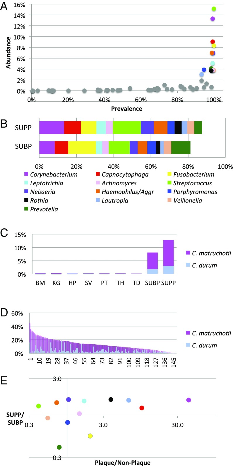
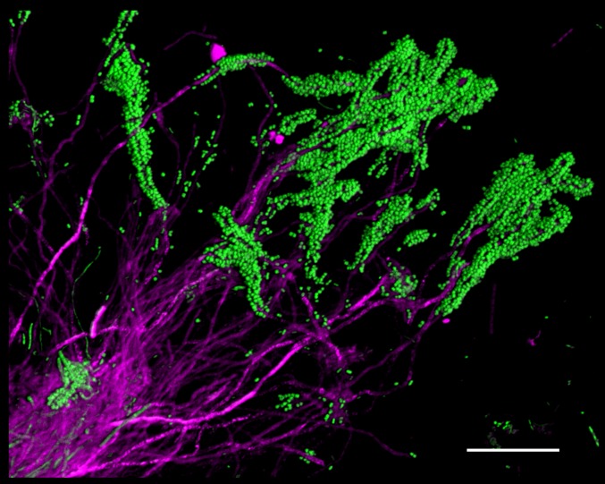
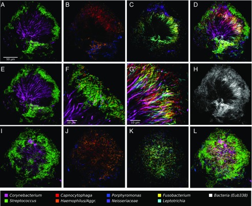
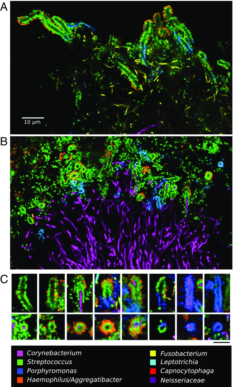
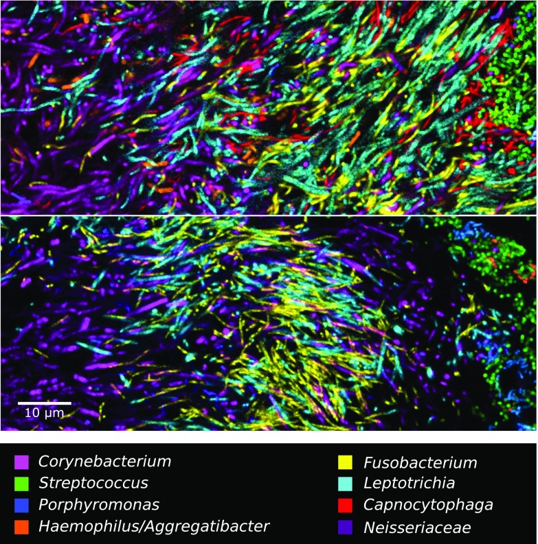
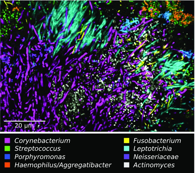
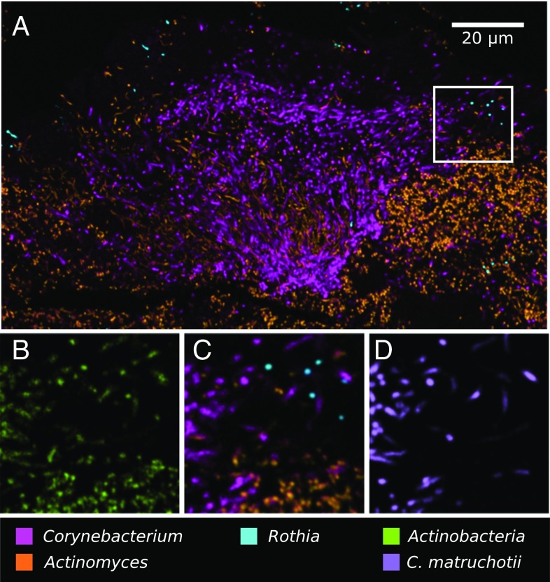
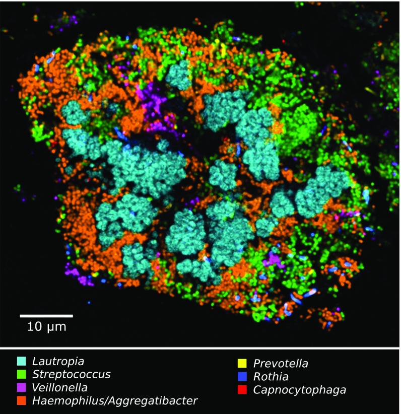
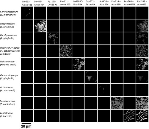
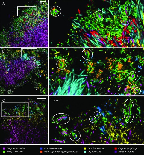
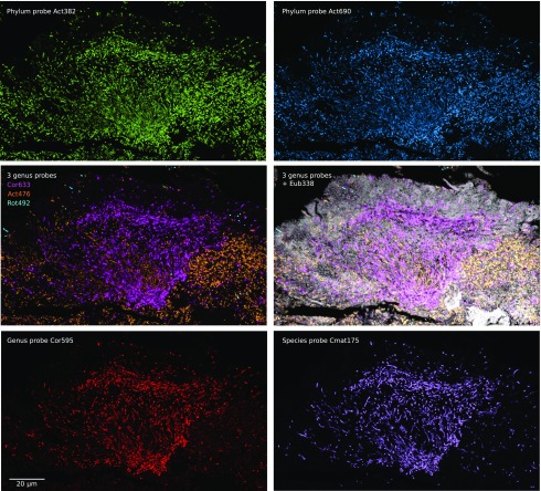
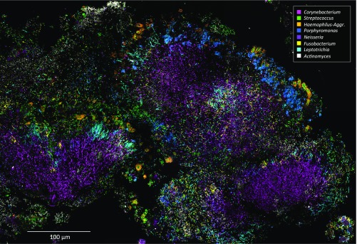
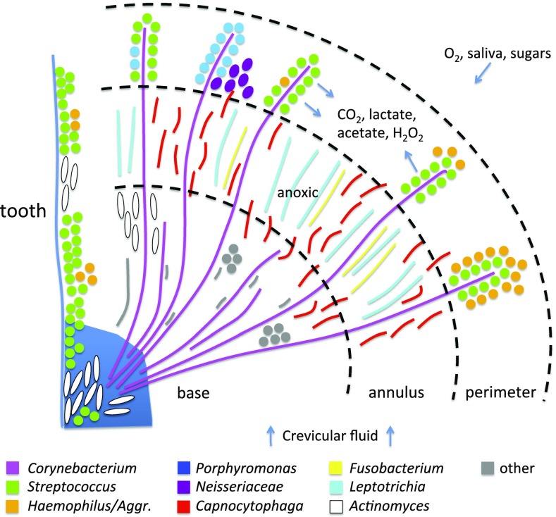
The spatial organization of complex natural microbiomes is critical to understanding the interactions of the individual taxa that comprise a community. Although the revolution in DNA sequencing has provided an abundance of genomic-level information, the biogeography of microbiomes is almost entirely uncharted at the micron scale. Using spectral imaging fluorescence in situ hybridization as guided by metagenomic sequence analysis, we have discovered a distinctive, multigenus consortium in the microbiome of supragingival dental plaque. The consortium consists of a radially arranged, nine-taxon structure organized around cells of filamentous corynebacteria. The consortium ranges in size from a few tens to a few hundreds of microns in radius and is spatially differentiated. Within the structure, individual taxa are localized at the micron scale in ways suggestive of their functional niche in the consortium. For example, anaerobic taxa tend to be in the interior, whereas facultative or obligate aerobes tend to be at the periphery of the consortium. Consumers and producers of certain metabolites, such as lactate, tend to be near each other. Based on our observations and the literature, we propose a model for plaque microbiome development and maintenance consistent with known metabolic, adherence, and environmental considerations. The consortium illustrates how complex structural organization can emerge from the micron-scale interactions of its constituent organisms. The understanding that plaque community organization is an emergent phenomenon offers a perspective that is general in nature and applicable to other microbiomes.

Keywords: biofilm; imaging; microbial ecology; microscopy.


Conflict of interest statement

The authors declare no conflict of interest.

Figures

Fig. 1.
Fig. 1.
Metagenomic sequence analysis points to Corynebacterium as a key taxon in supragingival plaque. (A) Prevalence abundance plot for supragingival plaque. (B) Cumulative abundance of genera in both supra- and subgingival plaque. Genera with greater than 3% abundance in SUPP, mean across 148 subjects, are indicated by colored dots in A and bar segments in B; B also shows the abundance of these genera in SUBP. Data are from the HMP (18) V3–V5 region of 16S rRNA, analyzed by oligotyping (19), and grouped by genus. (C) Corynebacterium is far more abundant in plaque than in other oral sites. Mean abundances of C. matruchotii and C. durum are shown for each oral site analyzed by oligotyping (19). BM, buccal mucosa; HP, hard palate; KG, keratinized gingiva; PT, palatine tonsils; SV, saliva; TD, tongue dorsum; TH, throat. (D) Corynebacterium is a major component of most plaque samples. Relative abundance of Corynebacterium in the HMP SUPP samples from 148 individuals (19). C. matruchotii is usually more abundant, but C. durum dominates some samples. (E) Habitat analysis identifies genera that are strongly characteristic of SUPP. The plaque to nonplaque ratio measures the relative abundance of each genus in two plaque sites compared with seven nonplaque sites sampled by the HMP [calculated as (mean SUBP + mean SUPP)/(mean BM + mean KG + mean HP + mean SV + mean PT + mean TH + mean TD)]. This ratio identifies Corynebacterium and Capnocytophaga as the taxa most preferentially abundant in plaque. The SUPP to SUBP ratio identifies these genera as relatively more abundant in SUPP than in SUBP. Colors in E are the same as those in A and B.
Fig. 2.
Fig. 2.
Corncob structures formed by Corynebacterium and cocci in plaque. Corynebacterium cells (magenta) are visible as long filaments, with cocci (green) bound to the tips of the filaments. Partially disrupted plaque was hybridized with a probe for Corynebacterium and a universal bacterial probe. Image was acquired using a Zeiss AxioImager 63× Plan-Apochromat 1.4 N.A. objective and Apotome structured illumination. (Scale bar: 20 μm.)
Fig. 3.
Fig. 3.
A hedgehog structure in plaque showing spatial organization of the plaque microbiome. Plaque was hybridized with a set of 10 probes each labeled with a different fluorophore. Each panel shows the superposition of several of these individual fluorophore channels. A–D and F–H show a single focal plane near the center of the structure, with two to three fluorophore channels shown in each of A–C and all nine specific probes superimposed in D. (E) Maximum intensity projection of three planes, representing a total of ∼2 μm of thickness, to visualize the continuity of Corynebacterium filaments from the center toward the edge of the structure. F is a detailed view of corncob structures. G is a detailed view of mixed filaments. H shows the fluorophore channel corresponding to the universal bacterial probe, showing that the specific probes (D) identify most of the cells that hybridize to the universal probe. I–L show a second focal plane near the periphery of the structure. Fluorophore channels shown correspond to the following genera in the figure: (A, E, and I) Corynebacterium and Streptococcus; (B and J) Capnocytophaga, Porphyromonas, and Haemophilus/Aggregatibacter; (C and K) Fusobacterium, Leptotrichia, and Neisseriaceae; (D and L) all nine specific probes; (F) Corynebacterium, Streptococcus, Porphyromonas, and Haemophilus/Aggregatibacter; (G) Corynebacterium, Fusobacterium, Leptotrichia, and Capnocytophaga; and (H) Bacteria. The plaque sample was fixed in 2% (wt/vol) paraformaldehyde, stored in 50% (vol/vol) ethanol, and spread onto the slide in 50% (vol/vol) ethanol in preparation for FISH.
Fig. 4.
Fig. 4.
Complex corncob structures in SUPP. (A and B) Clusters of corncobs at the perimeter of hedgehog structures. (A) Whole mount of plaque hybridized with probes for Corynebacterium, Fusobacterium, Streptococcus, Porphyromonas, and Haemophilus/Aggregatibacter. (B) Methacrylate-embedded section hybridized with probes for Corynebacterium, Streptococcus, Porphyromonas, and Haemophilus/Aggregatibacter. (C) Gallery of representative images showing types of corncobs frequently observed. (Scale bar: C, 5 μm.)
Fig. 5.
Fig. 5.
Filaments and rods of several genera intermingle at micron scales in an annulus of the hedgehog structure. The two images shown are from methacrylate-embedded, sectioned plaque from two different donors. Both samples were hybridized with probes for Corynebacterium, Fusobacterium, Leptotrichia, Streptococcus, Porphyromonas, Haemophilus/Aggregatibacter, and Neisseriaceae; the probe set in Upper also included a probe for Capnocytophaga.
Fig. 6.
Fig. 6.
Localization of Actinomyces within hedgehogs, in patches within the base region of hedgehogs, and adjacent to them.
Fig. 7.
Fig. 7.
Nested probing for species-level identification of Corynebacterium. Methacrylate-embedded, sectioned plaque was hybridized with a nested probe set targeting cells at the taxonomic levels of phylum, genus, and species. (A) Low-magnification image shows the three major oral genera of phylum Actinobacteria: Corynebacterium, Actinomyces, and Rothia. High-magnification views show (B) all Actinobacteria, (C) the three genera, and (D) C. matruchotii.
Fig. 8.
Fig. 8.
A cauliflower structure in plaque composed of Lautropia, Streptococcus, Haemophilus/Aggregatibacter, and Veillonella. Scattered cells of Prevotella, Rothia, and Capnocytophaga are also visible.
Fig. S1.
Fig. S1.
Probe set hybridizes as expected with pure cultures. The set of 10 probes, each labeled with a distinct fluorophore, was applied to pure cultures and subjected to imaging and linear unmixing under the same conditions used to image plaque samples. Each of nine taxon-specific probes hybridized with its target taxon and showed no significant hybridization to nontarget taxa. The near-universal probe Eub338 hybridized with all taxa, with variable intensity.
Fig. S2.
Fig. S2.
Corncob structures form around Corynebacterium and do not form around nearby Fusobacterium, Leptotrichia, or Capnocytophaga. A–C show methacrylate-embedded sections from three different donors. Rectangles indicate location of Insets; ovals highlight representative corncobs, each of which has a Corynebacterium core.
Fig. S3.
Fig. S3.
Nested probe set provides species-level identification of hedgehog Corynebacterium. Sample was hybridized with nine probes, each labeled with a different fluorophore, targeting cells at the level of kingdom, phylum, genus, and species. (Top) Two different probes targeting phylum Actinobacteria identify a consistent set of cells. Genus-level probes (Middle Left) identify these cells as the three genera Corynebacterium, Actinomyces, and Rothia and (Middle Right) are shown in the context of cells labeled with the universal probe Eub338 plus autofluorescence. (Bottom Left) A second genus-level probe validates the identity of Corynebacterium cells, and (Bottom Right) a species-level probe identifies them as Corynebacterium matruchotii.
Fig. S4.
Fig. S4.
Tile scan of a hedgehog structure in plaque. Image is a composite of seven fields of view showing a plaque sample with three adjacent hedgehogs.
Fig. 9.
Fig. 9.
Summary hypothesis for interpretation of hedgehog structures. Corynebacterium filaments bind to an existing biofilm containing Streptococcus and Actinomyces. At the distal tips of the Corynebacterium filaments, corncob structures form in which the filaments are surrounded by cocci, including Streptococcus and Porphyromonas, in direct contact with the Corynebacterium filament as well as Haemophilus/Aggregatibacter in contact with Streptococcus. Clusters of Neisseriaceae also occupy the periphery of the hedgehog. The Streptococcus cells create a microenvironment rich in CO2, lactate, and acetate, containing peroxide, and low in oxygen. Elongated filaments of Fusobacterium and Leptotrichia proliferate in this low-oxygen, high-CO2 environment in an annulus just proximal to the corncob-containing peripheral shell of the hedgehog. The CO2-requiring Capnocytophaga also proliferates abundantly in and around this annulus. The base of the hedgehog is dominated by Corynebacterium filaments and thinly populated by additional rods, filaments, and/or cocci.

Comment in

Similar articles

Cited by

References

    1. Diaz PI, Zilm PS, Rogers AH. Fusobacterium nucleatum supports the growth of Porphyromonas gingivalis in oxygenated and carbon-dioxide-depleted environments. Microbiology. 2002;148(Pt 2):467–472. - PubMed
    1. Mahowald MA, et al. Characterizing a model human gut microbiota composed of members of its two dominant bacterial phyla. Proc Natl Acad Sci USA. 2009;106(14):5859–5864. - PMC - PubMed
    1. Traxler MF, Watrous JD, Alexandrov T, Dorrestein PC, Kolter R. Interspecies interactions stimulate diversification of the Streptomyces coelicolor secreted metabolome. MBio. 2013;4(4):e00459-13. - PMC - PubMed
    1. Jakubovics NS. Intermicrobial interactions as a driver for community composition and stratification of oral biofilms. J Mol Biol. 2015;427(23):3662–3675. - PubMed
    1. Wright CJ, et al. Microbial interactions in building of communities. Mol Oral Microbiol. 2013;28(2):83–101. - PMC - PubMed

Publication types