. 2016 Feb 9;113(6):1534-9.
doi: 10.1073/pnas.1517517113. Epub 2016 Jan 25.

Biologic-free mechanically induced muscle regeneration

Affiliations

Biologic-free mechanically induced muscle regeneration

Christine A Cezar et al. Proc Natl Acad Sci U S A. .

Abstract

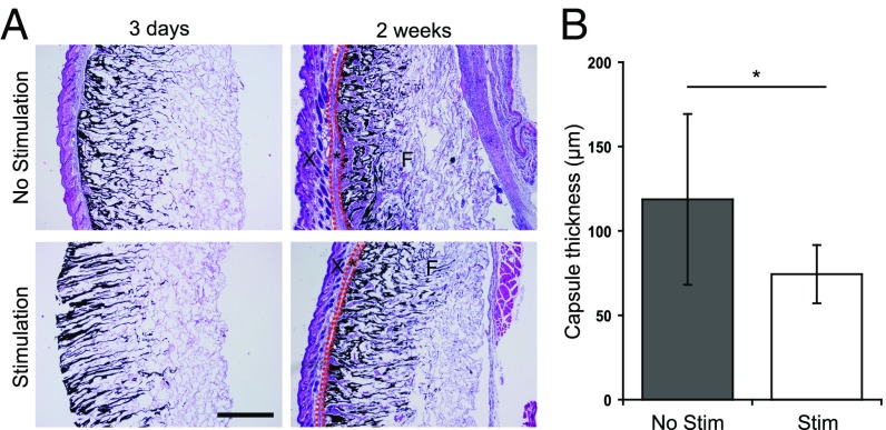
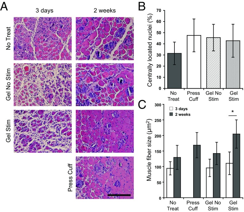
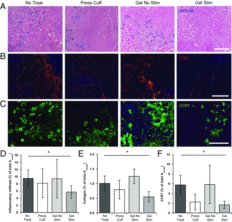
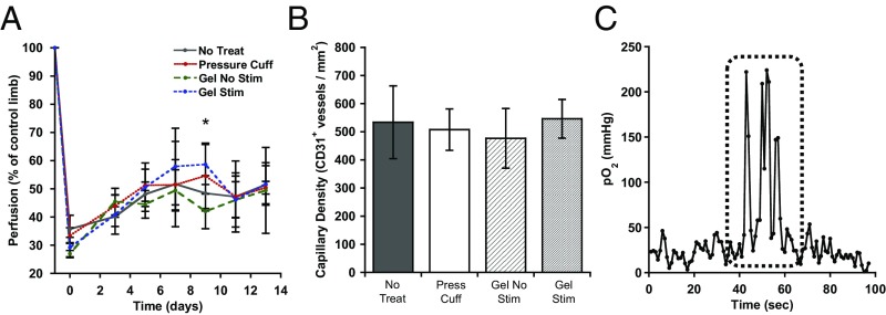
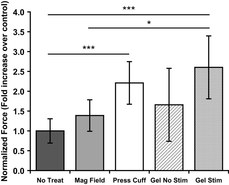
Severe skeletal muscle injuries are common and can lead to extensive fibrosis, scarring, and loss of function. Clinically, no therapeutic intervention exists that allows for a full functional restoration. As a result, both drug and cellular therapies are being widely investigated for treatment of muscle injury. Because muscle is known to respond to mechanical loading, we investigated instead whether a material system capable of massage-like compressions could promote regeneration. Magnetic actuation of biphasic ferrogel scaffolds implanted at the site of muscle injury resulted in uniform cyclic compressions that led to reduced fibrous capsule formation around the implant, as well as reduced fibrosis and inflammation in the injured muscle. In contrast, no significant effect of ferrogel actuation on muscle vascularization or perfusion was found. Strikingly, ferrogel-driven mechanical compressions led to enhanced muscle regeneration and a ∼threefold increase in maximum contractile force of the treated muscle at 2 wk compared with no-treatment controls. Although this study focuses on the repair of severely injured skeletal muscle, magnetically stimulated bioagent-free ferrogels may find broad utility in the field of regenerative medicine.

Keywords: fibrous capsule; immunomodulation; magnetic hydrogel; massage-mimetic; mechano-therapy.


Conflict of interest statement

The authors declare no conflict of interest.

Figures

Fig. 1.
Fig. 1.
Biphasic ferrogels and pressure cuffs generate cyclic mechanical compressions. (A) Experimental design showing injury, implant, and stimulation profile. (B) Schematic of biphasic ferrogel implant in mouse hind limb depicting orientation of ferrogel relative to skin, muscle tissue, and magnet (Left). Pressure profile of biphasic ferrogel undergoing repeated magnetic stimulations (Right). (C) Schematic of pressure cuff on mouse hind limb depicting orientation of balloon and polycarbonate cuff relative to skin and muscle tissue (Left). Pressure profile of balloon cuff undergoing repeated inflations and deflations (Right).
Fig. S1.
Fig. S1.
Biphasic ferrogels exhibit fatigue resistance. Percentage decrease in biphasic ferrogel (A) Young's modulus and (B) toughness at 50% strain after 8,400 cyclic compressions to 50% strain, compared with the values calculated from the first cycle of compression. Values represent the mean and SD (n = 4).
Fig. 2.
Fig. 2.
Magnetic stimulation of ferrogel implants decreases fibrous capsule thickness. (A) Cross-sections of biphasic ferrogels stained with H&E at 3 d and 2 wk after implantation. Skin (X), fibrous capsule (*), and ferrogels (F) are indicated. It is important to note that significant fibrous capsule formation was not observed at 3 d in either ferrogel condition, and surrounding tissues were often lost during processing. (B) Quantified fibrous capsule thickness of nonstimulated and stimulated biphasic ferrogels after 2 wk of implantation. Fibrous capsule boundaries are marked with red dashed lines in A. (Scale bar, 500 μm.) Data were compared using a two-tailed unpaired Student's test with Welch's correction (n = 9; *P < 0.05). Error bars represent SDs.
Fig. 3.
Fig. 3.
Ferrogel stimulation leads to improved muscle regeneration. (A) Histological cross-sections of tibialis anterior muscles stained with H&E 3 d and 2 wk after no treatment (No Treat), treatment with a pressure cuff (Press Cuff), treatment with a nonstimulated biphasic ferrogel (Gel No Stim), or treatment with a stimulated biphasic ferrogel (Gel Stim). (Scale bar, 100 μm.) (B) Quantification of myofibers residing in the defect containing centrally located nuclei 2 wk posttreatment. Values are expressed as a percentage of the total number of myofibers in the defect. (C) Quantified mean muscle fiber size in the defect area 3 d and 2 wk posttreatment. Data were compared using ANOVA with Bonferroni's post hoc test (n = 5; *P < 0.05). Error bars represent SDs.
Fig. 4.
Fig. 4.
Ferrogel stimulation decreases inflammation and fibrosis. (A and D) Representative images and quantification of the inflammatory infiltrate (INFLAM) in histological cross-sections of tibialis anterior muscles stained with H&E 2 wk posttreatment. (B and E) Representative images and quantification of tissue collagen (COLL) from picrosirius red stained cross-sections 2 wk posttreatment. (C and F) Representative images and quantification of M1 macrophages from CCR7-stained cross-sections 2 wk posttreatment. All values are expressed as a percentage of the total cross-section area (Across) of the tissue section. (Scale bars, 200 μm.) Data were compared using ANOVA with Dunnett's post hoc test (n = 5–10; *P < 0.05). Error bars represent SDs.
Fig. 5.
Fig. 5.
Intramuscular oxygen concentration increases during ferrogel stimulation. (A) Quantified perfusion of injured hind limbs normalized to contralateral controls, as measured by Laser Doppler perfusion imaging. *A difference between the stimulated and nonstimulated biphasic ferrogel conditions appeared at day 9. (B) Quantified capillary density in injured muscle, as assessed by CD31+ staining 2 wk posttreatment. (C) Representative oxygen probe trace with stimulation period marked by a dashed line. Data were compared using ANOVA with Bonferroni's post hoc test (n = 5; *P < 0.05). Error bars represent SDs.
Fig. 6.
Fig. 6.
Cyclic mechanical compressions enhance functional muscle regeneration. Maximum contractile force after tetanic stimulation of injured muscles 2 wk after no treatment (No Treat), treatment with a magnetic field only (Mag Field), treatment with a pressure cuff (Press Cuff), treatment with a nonstimulated biphasic ferrogel (Gel No Stim), or treatment with a stimulated biphasic ferrogel (Gel Stim). Force measurements were normalized to muscle wet weight. Data were compared using ANOVA with Bonferroni’s post hoc test (n = 5–10; *P < 0.05; ***P < 0.001).

References

    1. Juhas M, Bursac N. Engineering skeletal muscle repair. Curr Opin Biotechnol. 2013;24(5):880–886. - PMC - PubMed
    1. Turner NJ, Badylak SF. Regeneration of skeletal muscle. Cell Tissue Res. 2012;347(3):759–774. - PubMed
    1. Järvinen TAHJ, Järvinen TLN, Kääriäinen M, Kalimo H, Järvinen M. Muscle injuries: Biology and treatment. Am J Sports Med. 2005;33(5):745–764. - PubMed
    1. Rossi CA, Pozzobon M, De Coppi P. Advances in musculoskeletal tissue engineering: Moving towards therapy. Organogenesis. 2010;6(3):167–172. - PMC - PubMed
    1. Ma CH, et al. Reconstruction of upper extremity large soft-tissue defects using pedicled latissimus dorsi muscle flaps--technique illustration and clinical outcomes. Injury. 2008;39(Suppl 4):67–74. - PubMed

Publication types

MeSH terms