. 2016 Apr 1;34(10):1112-21.
doi: 10.1200/JCO.2015.64.5929. Epub 2016 Jan 25.

Allogeneic T Cells That Express an Anti-CD19 Chimeric Antigen Receptor Induce Remissions of B-Cell Malignancies That Progress After Allogeneic Hematopoietic Stem-Cell Transplantation Without Causing Graft-Versus-Host Disease

Affiliations

Allogeneic T Cells That Express an Anti-CD19 Chimeric Antigen Receptor Induce Remissions of B-Cell Malignancies That Progress After Allogeneic Hematopoietic Stem-Cell Transplantation Without Causing Graft-Versus-Host Disease

Jennifer N Brudno et al. J Clin Oncol. .

Abstract

Purpose: Progressive malignancy is the leading cause of death after allogeneic hematopoietic stem-cell transplantation (alloHSCT). After alloHSCT, B-cell malignancies often are treated with unmanipulated donor lymphocyte infusions (DLIs) from the transplant donor. DLIs frequently are not effective at eradicating malignancy and often cause graft-versus-host disease, a potentially lethal immune response against normal recipient tissues.

Methods: We conducted a clinical trial of allogeneic T cells genetically engineered to express a chimeric antigen receptor (CAR) targeting the B-cell antigen CD19. Patients with B-cell malignancies that had progressed after alloHSCT received a single infusion of CAR T cells. No chemotherapy or other therapies were administered. The T cells were obtained from each recipient's alloHSCT donor.

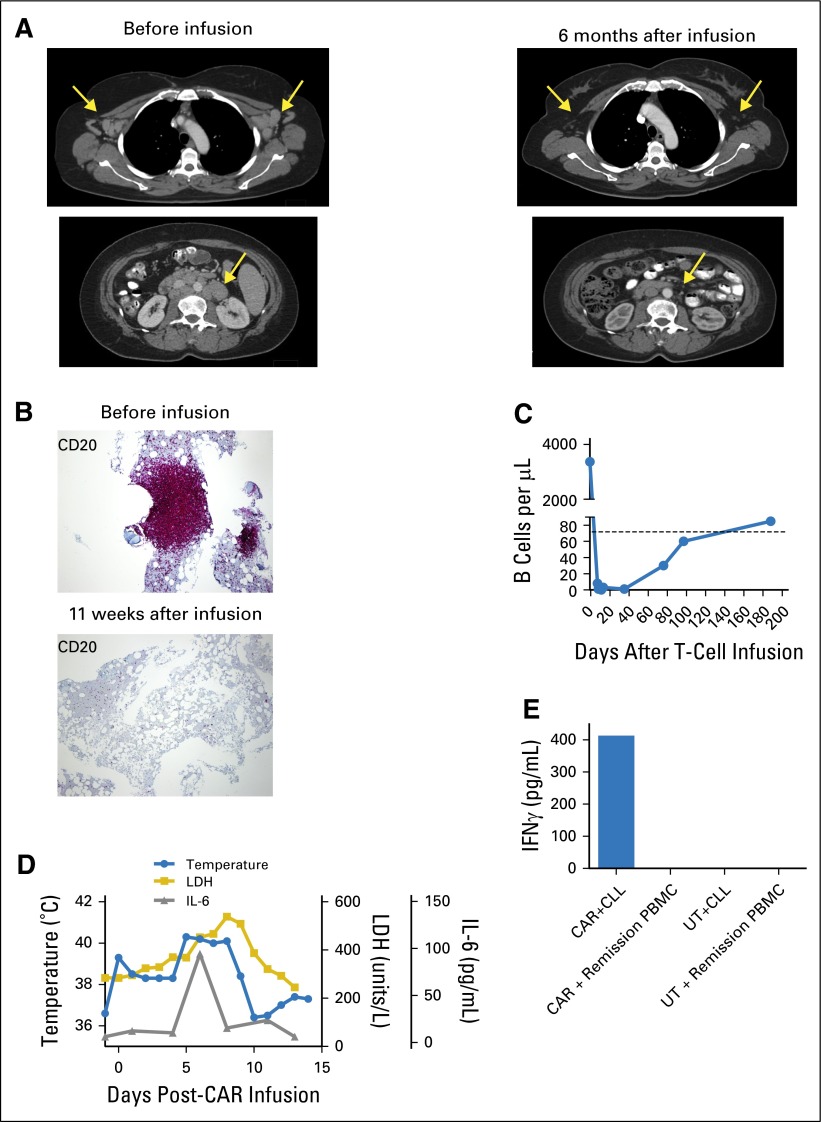
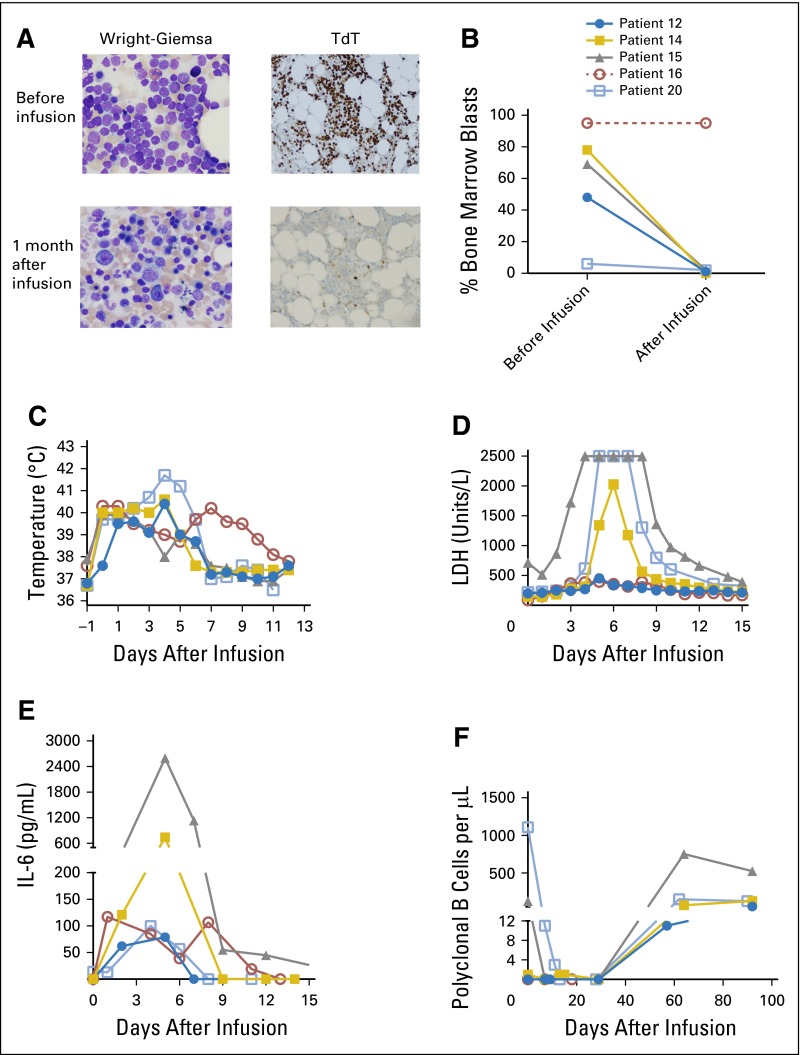
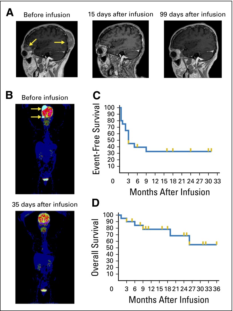
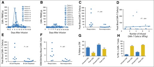
Results: Eight of 20 treated patients obtained remission, which included six complete remissions (CRs) and two partial remissions. The response rate was highest for acute lymphoblastic leukemia, with four of five patients obtaining minimal residual disease-negative CR. Responses also occurred in chronic lymphocytic leukemia and lymphoma. The longest ongoing CR was more than 30 months in a patient with chronic lymphocytic leukemia. New-onset acute graft-versus-host disease after CAR T-cell infusion developed in none of the patients. Toxicities included fever, tachycardia, and hypotension. Peak blood CAR T-cell levels were higher in patients who obtained remissions than in those who did not. Programmed cell death protein-1 expression was significantly elevated on CAR T cells after infusion. Presence of blood B cells before CAR T-cell infusion was associated with higher postinfusion CAR T-cell levels.

Conclusion: Allogeneic anti-CD19 CAR T cells can effectively treat B-cell malignancies that progress after alloHSCT. The findings point toward a future when antigen-specific T-cell therapies will play a central role in alloHSCT.


Conflict of interest statement

Authors’ disclosures of potential conflicts of interest are found in the article online at www.jco.org. Author contributions are found at the end of this article.

Figures

Fig 1.
Fig 1.
Rapid reduction of chronic lymphocytic leukemia (CLL) in lymph nodes, bone marrow, and blood after infusion of unrelated donor anti-CD19 chimeric antigen receptor (CAR19) T cells. (A) Computerized axial tomography scans demonstrate elimination of adenopathy (arrows) after CAR19 T-cell infusion. (B) CD20 immunohistochemical staining of bone marrow demonstrates eradication of bone marrow CLL (violet mass) after CAR19 T-cell infusion. (C) Before CAR19 T-cell infusion, patient 11 had a blood CD19+ B-cell count of 3,372 cells/μL (normal B-cell count, 81 to 493/μL). The B-cell count was elevated because of a large burden of CD19+ CLL cells. On day 11 after CAR19 T-cells infusion, the blood B-cell count was 0 cells/μL, which indicates a complete elimination of both CLL and normal B cells from the blood. Polyclonal B cells recovered to normal levels by 188 days after CAR19 T-cell infusion. (D) During the first 10 days after CAR19 T-cell infusion, serum lactate dehydrogenase (LDH), serum interleukin-6 (IL-6), and temperature were elevated. (E) Specific recognition of CLL cells by CAR19 T cells was demonstrated in vitro. T cells from patient 11’s unrelated donor were either transduced with the CAR19 vector or left untransduced. Both CAR19 T cells and untransduced donor T cells were then cultured in vitro with the patient’s CLL cells for 9 days to simulate in vivo exposure to CLL cells. After this culture period, the CAR19 T cells (CAR) recognized the patient’s CLL as shown by interferon γ (IFNγ) production in an enzyme-linked immunosorbent assay, whereas the untransduced T cells (UT) failed to recognize the CLL. Neither the CAR19 nor the untransduced donor T cells produced IFNγ in response to remission peripheral blood mononuclear cells (PBMCs) obtained from patient 11 after CAR19 T-cell infusion when CLL cells and normal B cells were absent from the blood.
Fig 2.
Fig 2.
Allogeneic anti-CD19 chimeric antigen receptor (CAR19) T cells are highly effective against acute lymphoblastic leukemia (ALL). (A) Patient 14 obtained a minimal residual disease–negative complete remission and had reconstitution of normal hematopoiesis after CAR19 T-cell infusion. Wright-Giemsa and terminal deoxynucleotidyl transferase (TdT) stains are shown. (B) The bone marrow blast percentages from before CAR19 T-cell infusion and 1 month after the infusion are shown. Blast percentages were determined morphologically on bone marrow aspirates. ALL in patient 16 did not respond to the CAR T-cell infusion as determined by peripheral blood counts, and no follow-up bone marrow biopsy was performed, so this patient’s postinfusion bone marrow blast percentage is reported as 95%, which was the same as that before infusion. Because no postinfusion bone marrow biopsy was performed on patient 16, the line for this patient is dashed. The symbols for each patient are defined in (B). The same symbols are used for each patient in (C), (D), (E), and (F). (C) Fever developed in all the patients with ALL after infusion of CAR19 T cells. The maximum daily temperature of each patient is shown. (D) Serum lactase dehydrogenase (LDH) levels increased in all patients with ALL during the time when they were experiencing clinical toxicity. The maximum measurable serum LDH concentration was 2,500 units/L, and patients 15 and 20 both had serum LDH concentrations of greater than 2,500 units/L on multiple days. (E) Serum interleukin-6 (IL-6) levels increased after CAR19 T-cell infusion. (F) In patients 15 and 20, normal polyclonal B cells were eradicated after CAR19 T-cell infusion. The other patients with ALL already had very low blood B-cell levels before CAR19 T-cell infusion. Recovery of polyclonal B cells was detected in four of five patients with ALL. Patient 16 did not recover polyclonal B cells by 29 days after infusion, and after that time, was not evaluable for B-cell recovery.
Fig 3.
Fig 3.
Rapid eradication of diffuse large–B-cell lymphoma after allogeneic CAR19 T-cell infusion. (A) Magnetic resonance images show rapid complete elimination of lymphoma masses (arrows). Images are from before treatment, 15 days after anti-CD19 chimeric antigen receptor (CAR19) T-cell infusion, and 99 days after CAR19 T-cell infusion. The remission continued over 6 months after infusion. (B) Positron emission tomography scan shows metabolically active lymphoma before CAR19 T-cell infusion (arrows). Thirty-five days after the CAR19 T-cell infusion, the scan showed no evidence of lymphoma. (C) The event-free survival of all 20 patients treated on the study is shown. Events were defined as progression of malignancy, receipt of any antimalignancy therapy after CAR19 T-cell infusion, or death from any cause. (D) The overall survival of all 20 patients treated on the study is shown. For (C) and (D), survival fractions were calculated by Kaplan-Meier method, and gold lines indicate censored patients.
Fig 4.
Fig 4.
High peak blood levels of anti-CD19 chimeric antigen receptor (CAR19) T cells were associated with remissions of malignancy. (A) The absolute numbers of blood T cells that contained the CAR gene were assessed by quantitative polymerase chain reaction (qPCR) before infusion and at multiple time points after infusion. Results for patients who obtained a response of either a complete remission (CR) or a partial remission (PR) are shown. (B) The absolute numbers of T cells that contained the CAR gene were assessed by qPCR for all patients with malignancies that did not respond to CAR19 T cells. Lack of response was defined as a response of stable disease (SD) or progressive disease (PD). (C) The peak numbers of blood CAR-positive (CAR+) T cells were higher in responders (patients obtaining CR or PR) than in nonresponders (patients with SD or PD). CAR+ T cells were measured by qPCR. P = .001 by Mann-Whitney test. (D) Linear regression analysis of the number of infused CAR+ T cells per kilogram for each patient versus the peak number of blood CAR+ T cells for each patient was performed. Blood CAR+ T cells were measured by qPCR. The peak CAR+ T-cell level was not predicted by the number of infused CAR+ T cells (R2 = 0.004). (E) Patients with blood B-lymphocyte counts above the lower limit of normal before CAR19 T-cell infusion (B-cell replete) had higher peak blood CAR+ T-cell levels than patients with low blood B-lymphocyte counts before CAR19 T-cell infusion (B-cell depleted). CAR+ T cells were measured by qPCR. The normal range for blood B lymphocytes is 81 to 493/mL. B lymphocytes were defined as CD19+ lymphocytes, which included both normal lymphocytes and chronic lymphocytic leukemia lymphocytes. The median blood B-cell count for B-cell–replete patients was 322/μL. The median blood B-cell count for B-cell–depleted patients was 1/μL. The groups were compared by Mann-Whitney test. For (C), (D), and (E), analysis was performed on all 20 treated patients. (F) Flow cytometry staining with a monoclonal antibody specific for CAR19 was performed. Cells were also stained for CD3, CD4, and CD8. The CD8:CD4 ratios of CD3+ CAR+ cells were calculated. Flow cytometry was performed on peripheral blood mononuclear cells (PBMCs) obtained 5 to 14 days after CAR T-cell infusion during the time of each patient’s peak CAR19 blood level. The fraction of CAR19 T cells that were CD8+ was higher for patients with responses of CR or PR (responders) than for patients with outcomes of SD or PD (nonresponders). The groups were compared by Mann-Whitney test. (G) Flow cytometry was performed on a sample of the infused T cells or on PBMCs from the time of each patient’s peak CAR19 T-cell level between 5 and 14 days after infusion. For this analysis, naïve T cells were defined as cells with a CD45RA+ C-C chemokine receptor type 7–positive (CCR7+) phenotype, and central memory (CM) T cells were defined as cells with a CD45RA CCR7+ phenotype. A substantial fraction of the CAR+ T cells had a naïve or CM phenotype at the time of infusion. For both CD8+ and CD4+ T cells, the fraction of CAR-expressing T cells with a naïve or CM phenotype decreased between the time of infusion and the time of peak blood levels of CAR19 T cells. The mean and SEM are shown for each category. The Wilcoxon matched pairs signed rank test was used to compare the fraction of naïve plus CM cells among the infused CAR-expressing T cells to the fraction of naïve plus CM cells among CAR-expressing T cells from the time of peak blood CAR T-cell levels (for both CD8+ and CD4+ T cells, P < .001). (H) Flow cytometry to detect programmed cell death protein-1 (PD-1) was performed on a sample of the infused T cells or on PBMCs from the time of peak CAR19 levels. The fraction of both CD8+ and CD4+ CAR+ T cells expressing PD-1 increased between the time of infusion and the time of peak blood CAR19 T-cell levels. The mean and SEM are shown for each category. By the Wilcoxon matched pairs signed rank test, P < .001 for both the CD8 and the CD4 comparisons. For (F), (G), and (H), analyses were performed on all 16 patients with detectable blood CAR19 T cells and available blood samples.

References

    1. Khouri IF, Champlin RE. Nonmyeloablative allogeneic stem cell transplantation for non-Hodgkin lymphoma. Cancer J. 2012;18:457–462. - PMC - PubMed
    1. Pavletic SZ, Kumar S, Mohty M, et al. NCI First International Workshop on the Biology, Prevention, and Treatment of Relapse after Allogeneic Hematopoietic Stem Cell Transplantation: Report from the Committee on the Epidemiology and Natural History of Relapse Following Allogeneic Cell Transplantation. Biol Blood Marrow Transplant. 2010;16:871–890. - PMC - PubMed
    1. van den Brink MR, Porter DL, Giralt S, et al. Relapse after allogeneic hematopoietic cell therapy. Biol Blood Marrow Transplant. 2010;16:S138–S145. - PMC - PubMed
    1. Spyridonidis A, Labopin M, Schmid C, et al. Immunotherapy Subcommittee of Acute Leukemia Working Party Outcomes and prognostic factors of adults with acute lymphoblastic leukemia who relapse after allogeneic hematopoietic cell transplantation. An analysis on behalf of the Acute Leukemia Working Party of EBMT. Leukemia. 2012;26:1211–1217. - PubMed
    1. Thomson KJ, Morris EC, Bloor A, et al. Favorable long-term survival after reduced-intensity allogeneic transplantation for multiple-relapse aggressive non-Hodgkin’s lymphoma. J Clin Oncol. 2009;27:426–432. - PubMed

Publication types

MeSH terms