

Anatomical variations of the celiac trunk and hepatic arterial system: an analysis using multidetector computed tomography angiography
- PMID: 26811552
- PMCID: PMC4725396
- DOI: 10.1590/0100-3984.2014.0100
Anatomical variations of the celiac trunk and hepatic arterial system: an analysis using multidetector computed tomography angiography
Abstract
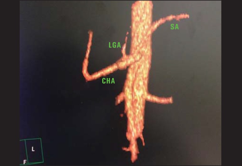
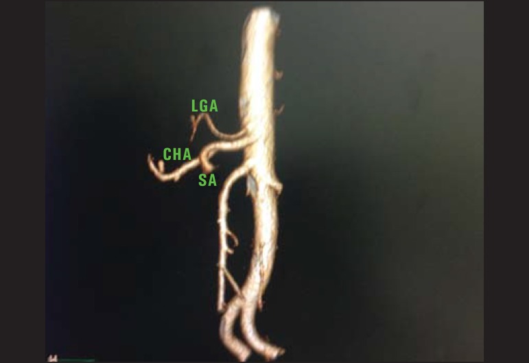
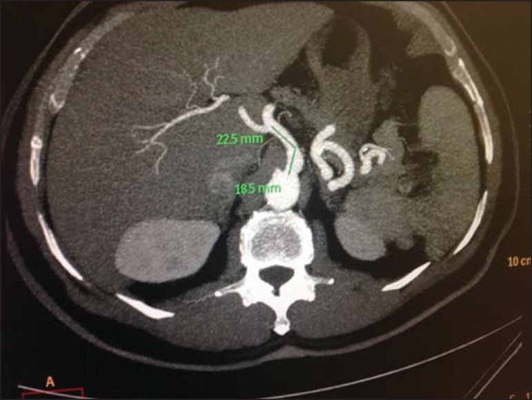
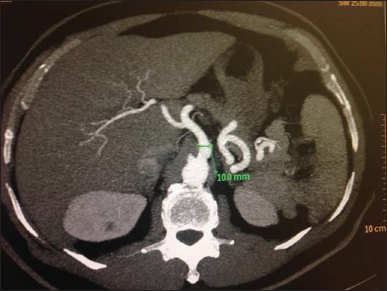
Objective: To analyze the prevalence of anatomical variations of celiac arterial trunk (CAT) branches and hepatic arterial system (HAS), as well as the CAT diameter, length and distance to the superior mesenteric artery.
Materials and methods: Retrospective, cross-sectional and predominantly descriptive study based on the analysis of multidetector computed tomography images of 60 patients.
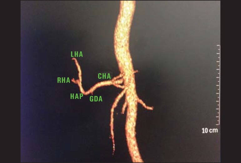
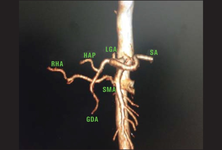
Results: The celiac trunk anatomy was normal in 90% of cases. Hepatosplenic trunk was found in 8.3% of patients, and hepatogastric trunk in 1.7%. Variation of the HAS was observed in 21.7% of cases, including anomalous location of the right hepatic artery in 8.3% of cases, and of the left hepatic artery, in 5%. Also, cases of joint relocation of right and left hepatic arteries, and trifurcation of the proper hepatic artery were observed, respectively, in 3 (5%) and 2 (3.3%) patients. Mean length and caliber of the CAT were 2.3 cm and 0.8 cm, respectively. Mean distance between CAT and superior mesenteric artery was 1.2 cm (standard deviation = 4.08). A significant correlation was observed between CAT diameter and length, and CAT diameter and distance to superior mesenteric artery.
Conclusion: The pattern of CAT variations and diameter corroborate the majority of the literature data. However, this does not happen in relation to the HAS.
Objetivo: Analisar a prevalência de variações anatômicas da ramificação do tronco arterial celíaco (TAC) e do sistema arterial hepático (SAH), o diâmetro e comprimento do TAC e sua distância para a artéria mesentérica superior.
Materiais e métodos: Estudo retrospectivo, transversal, predominantemente descritivo, baseado na análise de imagens de tomografia computadorizada de 60 pacientes.
Resultados: A anatomia do TAC foi normal em 90% dos casos. Cinco (8,3%) pacientes apresentaram o tronco hepatoesplênico e um (1,7%) apresentou o tronco hepatogástrico. O SAH variou em 21,7% dos casos. Desses, 8,3% foram na localização anômala da artéria hepática direita e 5% da artéria hepática esquerda. Ainda foram encontrados 3 (5%) casos de relocalização conjunta da artéria hepática direita e artéria hepática esquerda e 2 (3,3%) de trifurcação da artéria hepática própria. A média de comprimento e o calibre médio do TAC foram, respectivamente, 2,33 cm e 0,8 cm. A distância média entre o TAC e a artéria mesentérica superior foi 1,2 cm, com desviopadrão de 4,08. Houve correlação significativa entre diâmetro e comprimento do TAC, e diâmetro do TAC e distância deste para a artéria mesentérica superior.
Conclusão: O padrão de variação do TAC e seu diâmetro corroboram a maioria dos dados da literatura, embora o mesmo não tenha ocorrido em relação ao SAH.
Keywords: Anatomy; Celiac trunk; Hepatic artery; Multidetector computed tomography.
Figures
Similar articles
-
Anatomical Variants Of Celiac Trunk, Hepatic And Renal Arteries In A Population Of Developing Country Using Multidetector Computed Tomography Angiography.J Ayub Med Coll Abbottabad. 2017 Jul-Sep;29(3):450-454. J Ayub Med Coll Abbottabad. 2017. PMID: 29076681
-
Celiac Trunk and Hepatic Arteries: Anatomical Variations of Liver Arterial Supply as Detected with Multidetector Computed Tomography in 1,520 Patients and its Clinical Importance.Clin Anat. 2020 Oct;33(7):1091-1101. doi: 10.1002/ca.23511. Epub 2019 Nov 19. Clin Anat. 2020. PMID: 31688959
-
Anatomical variation of celiac axis, superior mesenteric artery, and hepatic artery: Evaluation with multidetector computed tomography angiography.J Res Med Sci. 2016 Dec 26;21:129. doi: 10.4103/1735-1995.196611. eCollection 2016. J Res Med Sci. 2016. PMID: 28331515 Free PMC article.
-
A morphometric study of the celiac trunk and review of the literature.Clin Anat. 2013 Sep;26(6):741-50. doi: 10.1002/ca.22136. Epub 2012 Aug 7. Clin Anat. 2013. PMID: 22886953 Review.
-
Variations of the celiac trunk investigated by multidetector computed tomography: Systematic review and meta-analysis with clinical correlations.Clin Anat. 2020 Nov;33(8):1249-1262. doi: 10.1002/ca.23576. Epub 2020 Feb 18. Clin Anat. 2020. PMID: 32012339
Cited by
-
Unusual development of the celiac trunk and its clinical significance.J Vasc Bras. 2021 Mar 15;20:e20200032. doi: 10.1590/1677-5449.200032. J Vasc Bras. 2021. PMID: 34093673 Free PMC article.
-
A rare anastomosis between the common hepatic artery and the superior mesenteric artery: a case report.Surg Radiol Anat. 2017 Oct;39(10):1175-1179. doi: 10.1007/s00276-017-1859-2. Epub 2017 Apr 21. Surg Radiol Anat. 2017. PMID: 28432408 Free PMC article.
-
Exploring celiac trunk parameters in median arcuate ligament syndrome: A CT study.Surg Radiol Anat. 2024 Jun;46(6):805-810. doi: 10.1007/s00276-024-03352-7. Epub 2024 Apr 15. Surg Radiol Anat. 2024. PMID: 38622333
-
A Case of Unusual Vascularization of Upper Abdominal Cavity' Organs.Case Rep Vasc Med. 2018 Oct 31;2018:5738037. doi: 10.1155/2018/5738037. eCollection 2018. Case Rep Vasc Med. 2018. PMID: 30515342 Free PMC article.
-
Anatomic Variations of the Hepatic Artery in 5625 Patients.Radiol Cardiothorac Imaging. 2021 Aug 19;3(4):e210007. doi: 10.1148/ryct.2021210007. eCollection 2021 Aug. Radiol Cardiothorac Imaging. 2021. PMID: 34498005 Free PMC article.
References
-
- Malnar D, Klasan GS, Miletic D, et al. Properties of the celiac trunk - anatomical study. Coll Antropol. 2010;34:917–921. - PubMed
-
- Chen H, Yano R, Emura S, et al. Anatomic variation of the celiac trunk with special reference to hepatic artery patterns. Ann Anat. 2009;191:399–407. - PubMed
-
- Prakash, Rajini T, Mokhasi V, et al. Coeliac trunk and its branches: anatomical variations and clinical implications. Singapore Med J. 2012;53:329–331. - PubMed
-
- Matusz P, Miclaus GD, Ples H, et al. Absence of the celiac trunk: case report using MDCT angiography. Surg Radiol Anat. 2012;34:959–963. - PubMed
-
- Mburu KS, Alexander OJ, Hassan S, et al. Variations in the branching pattern of the celiac trunk in a Kenyan population. Int J Morphol. 2010;28:199–204.
LinkOut - more resources
Full Text Sources
Other Literature Sources
Miscellaneous