Review
. 2015 Nov-Dec;48(6):373-80.
doi: 10.1590/0100-3984.2014.0017.

Chest magnetic resonance imaging: a protocol suggestion

Affiliations
Review

Chest magnetic resonance imaging: a protocol suggestion

Bruno Hochhegger et al. Radiol Bras. 2015 Nov-Dec.

Abstract

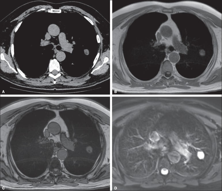
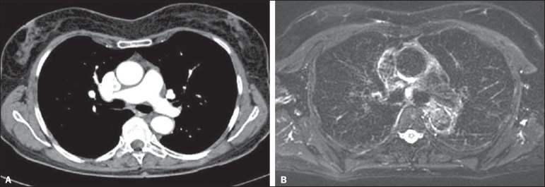
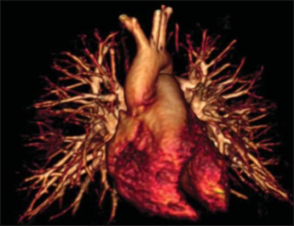
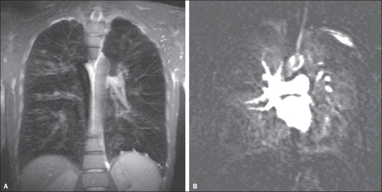
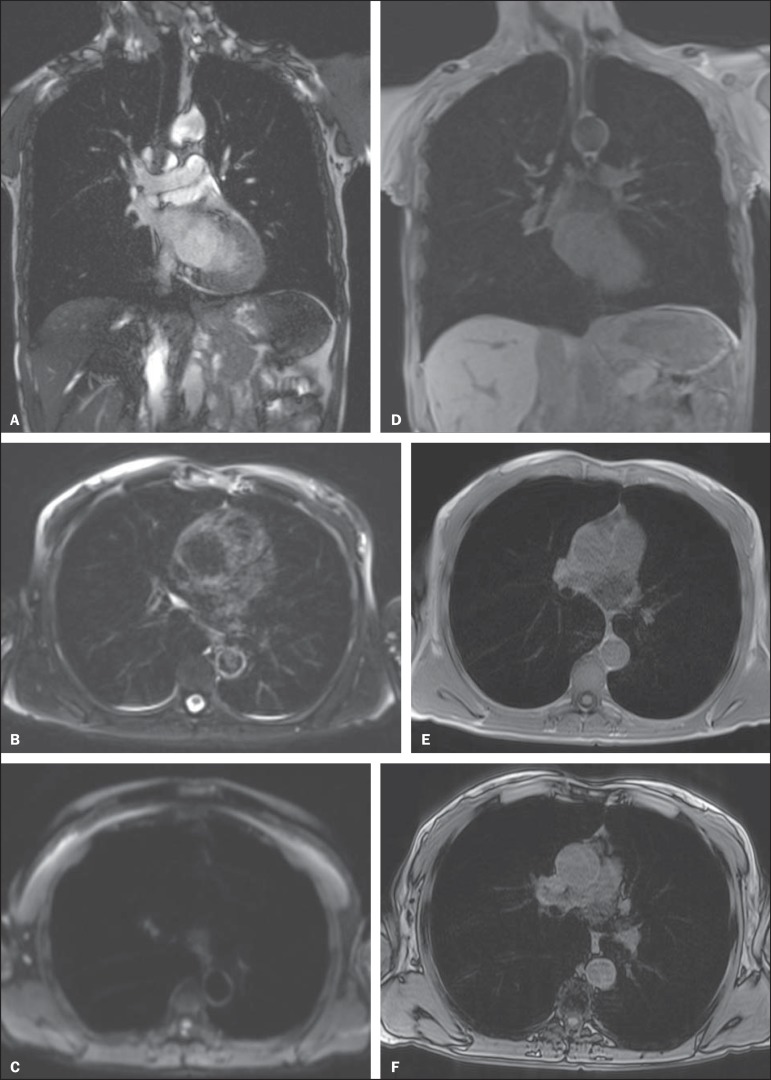
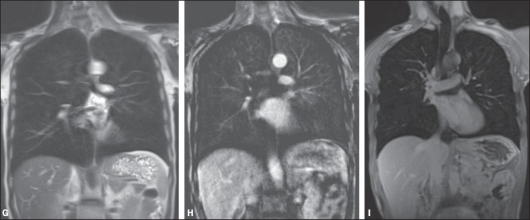
In the recent years, with the development of ultrafast sequences, magnetic resonance imaging (MRI) has been established as a valuable diagnostic modality in body imaging. Because of improvements in speed and image quality, MRI is now ready for routine clinical use also in the study of pulmonary diseases. The main advantage of MRI of the lungs is its unique combination of morphological and functional assessment in a single imaging session. In this article, the authors review most technical aspects and suggest a protocol for performing chest MRI. The authors also describe the three major clinical indications for MRI of the lungs: staging of lung tumors; evaluation of pulmonary vascular diseases; and investigation of pulmonary abnormalities in patients who should not be exposed to radiation.

Keywords: Chest; Lung; Magnetic resonance imaging; Protocol; Sequences.


Figures

Figure 1
Figure 1
A 56-year-old patient with pancreatic cancer. A: Axial CT image showing low attenuation areas inside the nodule. This nodule had mean -33 HU attenuation. In-phase (B) and opposed-phase (C) images showing signal loss in the nodule, suggesting hamartoma. D: T2-weighted sequence showing high signal intensity of the nodule.
Figure 2
Figure 2
A: Axial CT image showing a 8 mm lymph node in the subcarinal station. B: Axial T2-weighted image with fat saturation showing a high signal intensity in this lymph node, suggesting metastatic disease.
Figure 3
Figure 3
3D volume rendering of MR angiography showing subsegmental resolution.
Figure 4
Figure 4
32 year-old patient with cystic fibrosis. A: Coronal T1-weighted gradient echo sequence (VIBE) with 2 mm slice thickness. Note the presence of bronchiectasis with mucoid impaction. B: Pulmonary perfusion shows multiple perfusion defects better characterizing the disease severity.
Figure 5
Figure 5
A: Axial HRCT image showing homogeneous, segmental ground glass opacities in the pulmonary cortex. B: Axial T2-weighted image clearly demonstrates the lesion.
None
(A-F). Image examples for the suggested protocol. A: Coronal T2-weighted FSE TrueFisp sequence (4 mm). B: Axial, T2-weighted sequence Blade axial (4 mm). C: Axial, diffusion-weighted image (5 mm). D: Coronal, T1-weighted sequence VIBE with fat suppression (4 mm). E,F: T1-weighted sequence in- and out-of-phase.
None
(G–I): Image examples for the suggested protocol. G: Coronal, T2-weighted HASTE sequence. H: Coronal, T1-weighted perfusion sequence. I: T1-weighted sequence VIBE with fat suppression.

References

    1. Wild JM, Marshall H, Bock M, et al. MRI of the lung (1/3): methods. Insights Imaging. 2012;3:345–353. - PMC - PubMed
    1. Biederer J, Beer M, Hirsch W, et al. MRI of the lung (2/3). Why... when... how? Insights Imaging. 2012;3:355–371. - PMC - PubMed
    1. Koenigkam Santos M, Elias J, Júnior, Mauad FM, et al. Ressonância magnética do tórax: aplicações tradicionais e novas, com ênfase em pneumologia. J Bras Pneumol. 2011;37:242–258. - PubMed
    1. Koyama H, Ohno Y, Kono A, et al. Quantitative and qualitative assessment of non-contrast-enhanced pulmonary MR imaging for management of pulmonary nodules in 161 subjects. Eur Radiol. 2008;18:2120–2131. - PubMed
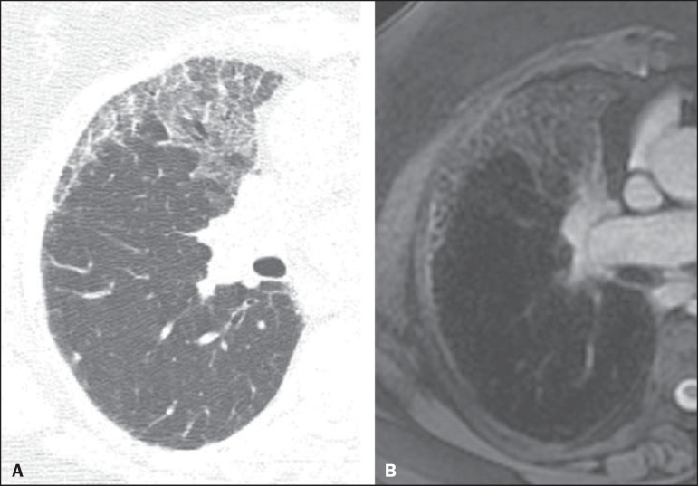
    1. Hochhegger B, Marchiori E, dos Reis DQ, et al. Chemical-shift MRI of pulmonary hamartomas: initial experience using a modified technique to assess nodule fat. AJR Am J Roentgenol. 2012;199:W331–W334. - PubMed

LinkOut - more resources