

Buried bumper syndrome: A complication of percutaneous endoscopic gastrostomy
- PMID: 26811611
- PMCID: PMC4716063
- DOI: 10.3748/wjg.v22.i2.618
Buried bumper syndrome: A complication of percutaneous endoscopic gastrostomy
Abstract
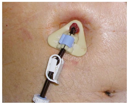
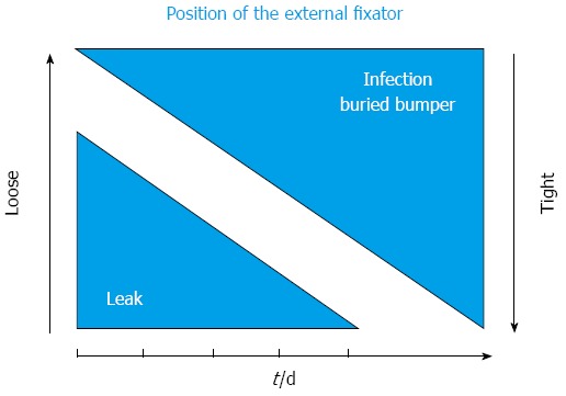
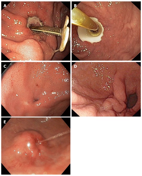
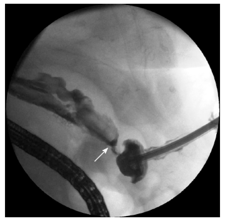
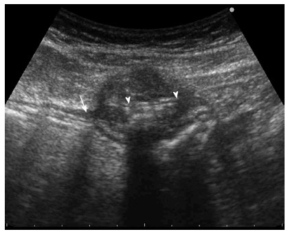
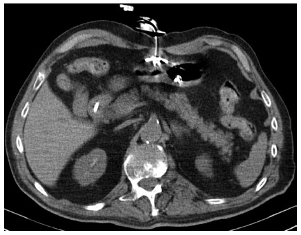
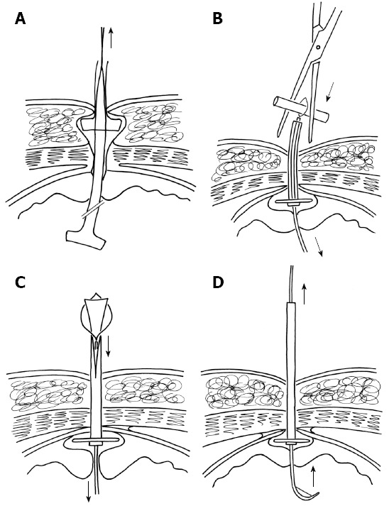
Percutaneous endoscopic gastrostomy (PEG) is a widely used method of nutrition delivery for patients with long-term insufficiency of oral intake. The PEG complication rate varies from 0.4% to 22.5% of cases, with minor complications being three times more frequent. Buried bumper syndrome (BBS) is a severe complication of this method, in which the internal fixation device migrates alongside the tract of the stoma outside the stomach. Excessive compression of tissue between the external and internal fixation device of the gastrostomy tube is considered the main etiological factor leading to BBS. Incidence of BBS is estimated at around 1% (0.3%-2.4%). Inability to insert, loss of patency and leakage around the PEG tube are considered to be a typical symptomatic triad. Gastroscopy is indicated in all cases in which BBS is suspected. The depth of disc migration in relation to the lamina muscularis propria of the stomach is critical for further therapy and can be estimated by endoscopic or transabdominal ultrasound. BBS can be complicated by gastrointestinal bleeding, perforation, peritonitis, intra-abdominal and abdominal wall abscesses, or phlegmon, and these complications can lead to fatal outcomes. The most important preventive measure is adequate positioning of the external bolster. A conservative approach should be applied only in patients with high operative risk and dismal prognosis. Choice of the method of release is based on the type of the PEG set and depth of disc migration. A disc retained inside the stomach and completely covered by the overgrowing tissue can be released using some type of endoscopic dissection technique (needle knife, argon plasma coagulation, or papillotome through the cannula). Proper patient selection and dissection of the overgrowing tissue are the major determinants for successful endoscopic therapy. A disc localized out of the stomach (lamina muscularis propria) should be treated by a surgeon.
Keywords: Buried bumper syndrome; Complication; Endoscopy; Enteral nutrition; Percutaneous endoscopic gastrostomy.
Figures
Comment in
-
Novel endoscopic management of buried bumper syndrome in percutaneous endoscopic gastrostomy: The Olympus HookKnife.World J Gastroenterol. 2017 Sep 21;23(35):6546-6548. doi: 10.3748/wjg.v23.i35.6546. World J Gastroenterol. 2017. PMID: 29085204 Free PMC article.
Similar articles
-
How to improve success rates of endoscopic management for buried bumper syndrome.QJM. 2018 Jul 1;111(7):467-472. doi: 10.1093/qjmed/hcy081. QJM. 2018. PMID: 29660086
-
[Buried bumper syndrome: A new classification and therapy algorithm].Chirurg. 2015 Oct;86(10):963-9. doi: 10.1007/s00104-014-2973-x. Chirurg. 2015. PMID: 25645431 Review. German.
-
Acute buried bumper syndrome.South Med J. 2010 Dec;103(12):1256-8. doi: 10.1097/SMJ.0b013e3181fa73d0. South Med J. 2010. PMID: 21037520
-
The buried bumper syndrome: the usefulness of retrieval PEG tubes in its management.Turk J Gastroenterol. 2008 Mar;19(1):45-8. Turk J Gastroenterol. 2008. PMID: 18386240
-
Buried bumper syndrome: do we have enough evidence?Br J Community Nurs. 2018 Jul 1;23(Sup7):S28-S30. doi: 10.12968/bjcn.2018.23.Sup7.S28. Br J Community Nurs. 2018. PMID: 30011232 Review.
Cited by
-
Managing complications of percutaneous tracheostomy and gastrostomy.J Thorac Dis. 2021 Aug;13(8):5314-5330. doi: 10.21037/jtd-19-3716. J Thorac Dis. 2021. PMID: 34527368 Free PMC article. Review.
-
May chronic cough in chronic obstructive pulmonary disease be a contraindication of Percutaneous Endoscopic Gastrostomy placement: a case report.BMC Gastroenterol. 2021 Jan 21;21(1):31. doi: 10.1186/s12876-021-01603-0. BMC Gastroenterol. 2021. PMID: 33478385 Free PMC article.
-
Buried Bumper Syndrome: a rare complication during radical chemoradiotherapy for head and neck cancer.BMJ Case Rep. 2021 May 25;14(5):e238203. doi: 10.1136/bcr-2020-238203. BMJ Case Rep. 2021. PMID: 34035012 Free PMC article.
-
Percutaneous Endoscopic Gastrostomy: Procedure, Complications and Management.Brain Neurorehabil. 2022 Mar 28;15(1):e2. doi: 10.12786/bn.2022.15.e2. eCollection 2022 Mar. Brain Neurorehabil. 2022. PMID: 36743844 Free PMC article. Review.
-
Early Recognition and Diagnosis of Buried Bumper Syndrome: A Report of Three Cases.Surg J (N Y). 2019 Aug 22;5(3):e76-e81. doi: 10.1055/s-0039-1692148. eCollection 2019 Jul. Surg J (N Y). 2019. PMID: 31448333 Free PMC article.
References
-
- Itkin M, DeLegge MH, Fang JC, McClave SA, Kundu S, d’Othee BJ, Martinez-Salazar GM, Sacks D, Swan TL, Towbin RB, et al. Multidisciplinary practical guidelines for gastrointestinal access for enteral nutrition and decompression from the Society of Interventional Radiology and American Gastroenterological Association (AGA) Institute, with endorsement by Canadian Interventional Radiological Association (CIRA) and Cardiovascular and Interventional Radiological Society of Europe (CIRSE) Gastroenterology. 2011;141:742–765. - PubMed
-
- Kwon RS, Banerjee S, Desilets D, Diehl DL, Farraye FA, Kaul V, Mamula P, Pedrosa MC, Rodriguez SA, Varadarajulu S, et al. Enteral nutrition access devices. Gastrointest Endosc. 2010;72:236–248. - PubMed
-
- Löser C, Aschl G, Hébuterne X, Mathus-Vliegen EM, Muscaritoli M, Niv Y, Rollins H, Singer P, Skelly RH. ESPEN guidelines on artificial enteral nutrition--percutaneous endoscopic gastrostomy (PEG) Clin Nutr. 2005;24:848–861. - PubMed
-
- McClave SA, Chang WK. Complications of enteral access. Gastrointest Endosc. 2003;58:739–751. - PubMed
-
- Gauderer MW, Ponsky JL, Izant RJ. Gastrostomy without laparotomy: a percutaneous endoscopic technique. J Pediatr Surg. 1980;15:872–875. - PubMed
Publication types
MeSH terms
LinkOut - more resources
Full Text Sources
Other Literature Sources
