

Comment
doi: 10.1016/j.cub.2015.11.026.
Visual Plasticity: Blindsight Bridges Anatomy and Function in the Visual System
Affiliations
- PMID: 26811892
- PMCID: PMC5172419
- DOI: 10.1016/j.cub.2015.11.026
Item in Clipboard
Comment
Visual Plasticity: Blindsight Bridges Anatomy and Function in the Visual System
Curr Biol.
.
Abstract
Some people who are blind due to damage to their primary visual cortex, V1, can discriminate stimuli presented within their blind visual field. This residual function has been recently linked to a pathway that bypasses V1, and connects the thalamic lateral geniculate nucleus directly with the extrastriate cortical area MT.
Copyright © 2016 Elsevier Ltd. All rights reserved.
Figures
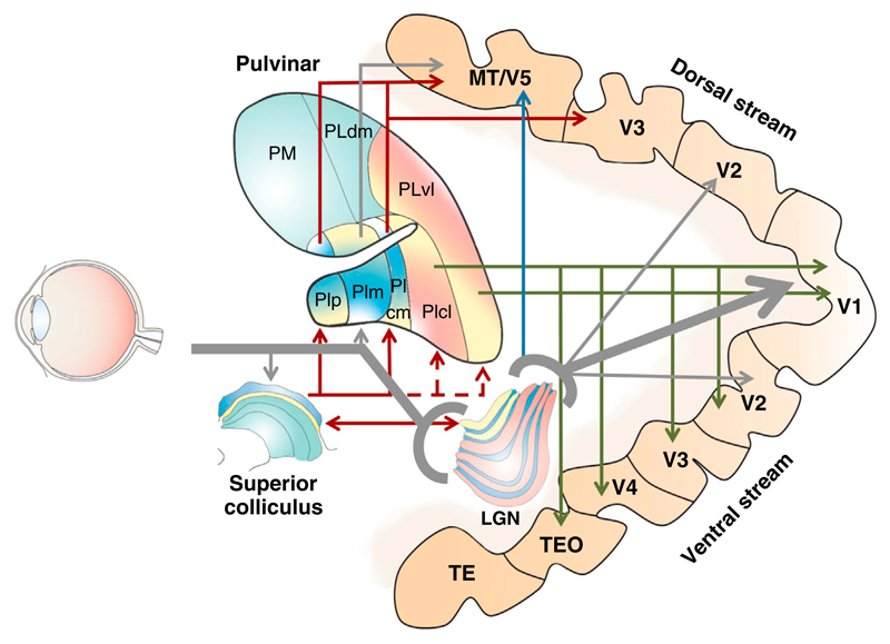
Gray arrows indicate direct projections for the eye, with thicker lines showing the major geniculo-striate pathway involving LGN and targeting V1. Red arrows indicate projections originating from the superior colliculus and reaching the dorsal stream cortical areas via the pulvinar, with dashed lines showing disputed input to subdivisions of the pulvinar. Green arrows indicate projections from pulvinar subnuclei to areas along the cortical ventral stream. The blue arrow indicates projections from the Koniocellular layers of LGN to area MT. In LGN and superior colliculus, yellow layers indicate Magnocellular, blue Koniocellular, and pink Parvocellular channels. In the pulvinar these pathways are not clearly segregated and shaded blue-yellow; pink-yellow colors indicate the conjoint presence of the respective channels in given subdivisions. Light green denotes areas of the superior colliculus and pulvinar not interesting for the present purposes. Abbreviations: PIcl, pulvinar inferior centro-lateral; PIcm, pulvinar inferior centro-medial; PIm, pulvinar inferior medial; PIp, pulvinar inferior posterior; PLdm, pulvinar lateral dorso-medial; PLvl, pulvinar lateral ventro-lateral; PM, pulvinar medial; TE, temporal inferior rostral; TEO, temporal inferior posterior.
Comment on
-
Human blindsight is mediated by an intact geniculo-extrastriate pathway.Elife. 2015 Oct 20;4:e08935. doi: 10.7554/eLife.08935. Elife. 2015. PMID: 26485034 Free PMC article.
References
-
- Fernel JF. De Naturali Parte Medicinae Libri Septem. Chapter 1. Paris: Simon de Colines; 1542.
-
- Riddoch G. Dissociation of visual perceptions due to occipital injuries, with especial reference to appreciation of movement. Brain. 1917;40:15–57.
-
- Weiskrantz L, Warrington EK, Sanders MD, Marshall J. Visual capacity in the hemianopic field following a restricted occipital ablation. Brain. 1974;97:709–728. - PubMed
-
- Leh SE, Johansen-Berg H, Ptito A. Unconscious vision: new insights into the neuronal correlate of blindsight using diffusion tractography. Brain. 2006;129:1822–1832. - PubMed
Publication types
MeSH terms
Grants and funding
LinkOut - more resources
Full Text Sources
Other Literature Sources
