. 2016 Jan 26;14(1):e1002361.
doi: 10.1371/journal.pbio.1002361. eCollection 2016 Jan.

Molecular Basis of Orb2 Amyloidogenesis and Blockade of Memory Consolidation

Affiliations

Molecular Basis of Orb2 Amyloidogenesis and Blockade of Memory Consolidation

Rubén Hervás et al. PLoS Biol. .

Abstract

Amyloids are ordered protein aggregates that are typically associated with neurodegenerative diseases and cognitive impairment. By contrast, the amyloid-like state of the neuronal RNA binding protein Orb2 in Drosophila was recently implicated in memory consolidation, but it remains unclear what features of this functional amyloid-like protein give rise to such diametrically opposed behaviour. Here, using an array of biophysical, cell biological and behavioural assays we have characterized the structural features of Orb2 from the monomer to the amyloid state. Surprisingly, we find that Orb2 shares many structural traits with pathological amyloids, including the intermediate toxic oligomeric species, which can be sequestered in vivo in hetero-oligomers by pathological amyloids. However, unlike pathological amyloids, Orb2 rapidly forms amyloids and its toxic intermediates are extremely transient, indicating that kinetic parameters differentiate this functional amyloid from pathological amyloids. We also observed that a well-known anti-amyloidogenic peptide interferes with long-term memory in Drosophila. These results provide structural insights into how the amyloid-like state of the Orb2 protein can stabilize memory and be nontoxic. They also provide insight into how amyloid-based diseases may affect memory processes.


Conflict of interest statement

The authors declare competing financial interest: MCV and RH are co-inventors on an international patent application (reference EP15382176.4) covering the results contained in this article. Any potential income generated by exploitation of the patent rights and received by their employer, the CSIC, shall be shared with these authors according to Spanish law and the regulations of the CSIC.

Figures

Fig 1
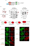
Fig 1. Orb2 forms self-propagating and canonical amyloids.
(A) Pictograms showing the domain organization of Orb2A and Orb2B isoforms. PLD, prion-like domain, in red; RRM, RNA-binding domains, in yellow; ZnF, zinc-finger motif, in purple. The N-terminal amino acids preceding the PLD of Orb2A (eight amino acids) and Orb2B (162 amino acids) are represented in green and blue, respectively. (B) In the presence of both Orb2A and Orb2B, ThT (Proteostat) shows enhanced fluorescence emission at 485 nm over time, a typical feature of amyloids, although the kinetics is much faster for Orb2A and Orb2B than other amyloids (see S1 Fig). (C) Concentration (μM) of the azo-dye CR bound to the amyloid component formed by Orb2. The data are represented as the mean ± standard error of the mean (SEM): ***p < 0.001 (One-way ANOVA and Tukey post-test). The inset shows a 63x image of an Orb2A amyloid under a polarized light microscope. (D) The CD spectra of soluble, full-length isoforms of Orb2 showed a α-helix-rich secondary structure. Deconvolution of the CD spectrum by using algorithms in DICHROWEB showed that Orb2A has 6% more α-helix and 5% less random coil conformations than Orb2B. (E) Relative abundance of secondary structural elements determined using FTIR. These distributions are significantly different from one another and from a non-amyloidogenic control protein concanavalin A control with a p-value of < 0.001 (chi-square test). (F) Aggregation of both isoforms over the incubation period monitored by turbidimetry at 405 nm. (G) Representative electron micrograph of oligomers (asterisks) and amyloid fibers (arrow) formed by Orb2A. Scale bar: 0.2 μm. (H) Schematic representation of the constructs used in the yeast prion assay (left panel). Orb2A-Sup35C efficiently switches to the prion state. Randomly selected clones were replica plated either in complete yeast extract peptone dextrose (YPD) media or in media lacking adenine. Only Orb2A produced a high frequency of red Ade- and white Ade+ colonies (right panel). NM: N-terminal and medial regions; C: C-terminal region. The underlying data for panels in this figure can be found in S1 Data.
Fig 2
Fig 2. Organization of the Orb2 PLD.
(A). Schematic representation of the location of the TEV protease recognition motif (ENLYFQG) inserted into Orb2. (B) EGFP-tagged Orb2A and Orb2B bearing TEV protease sites were expressed pan-neuronally, and immunopurified Orb2 was treated with TEV protease for 24 h. The left panel shows the schematic of the experiment. The cleaved C-terminus of different sizes derived from Orb2A are indicated with a bracket. The * indicates an EGFP reactive polypeptide most likely originated from degradation of the cleaved Orb2B370TEV C-terminus. (C) Schematic diagram of the epithelial fusion failure 1 (EFF-1) cell fusion “cytoduction” experiment in S2 cells. The expected outcomes in terms of the recruitment or nonrecruitment of oligomers are indicated. (D) Residues 88 to 162 are sufficient for recruitment into pre-existing oligomers. Full-length Orb2A-EGFP induced the Orb2A construct lacking the 88 N-terminal residues (Orb2AΔ1-88-Cherry) to oligomerize. Full-length Orb2A from D. willistoni failed to induce oligomerization of Orb2AΔ1-88-Cherry. (E) EGFP, Orb2B-EGFP, or Orb1-EGFP also failed to induce oligomerization of Orb2AΔ1-88-Cherry. Representative examples of fused cells are shown and the n for each experimental set is ≥10. Scale bar = 5 μm.
Fig 3
Fig 3. The F5 residue in Orb2A is critical for Orb2 amyloid-like oligomer formation and self-sustaining prion-like properties.
(A) Schematic representation of the experimental design and the positions analyzed by the indicated methods. (B) FRAP was used to measure the dynamic nature of the Orb2A-EGFP aggregates, showing that substitution of the 5th residue makes the aggregates more dynamic. (C) The F5Y mutation reduces the rate of amyloid formation, as gauged by ThT fluorescence. (D) FRET was used to measure the relative orientation and organization of the Orb2A proteins in the aggregate. For Fig 3B and 3D analysis, data are represented as mean ± SEM. We assumed statistical significance at *p < 0.05, **p < 0.01 and ***p < 0.001 (One-way ANOVA). (E) A single residue substitution in Orb2A, F5Y, dramatically reduced the ability to adopt the prion-like state. (F) Orb2A harbouring the F5Y mutation partitions less into the pellet fraction, and it forms less SDS-resistant oligomers than the wild type form. (G) Orb2AF5Y-EGFP did not induce oligomerization of Orb2A lacking the 88 N-terminal residues (Orb2AΔ1-88-Cherry). Scale bar: 5 μm. The underlying data for panels in this figure can be found in S1 Data.
Fig 4
Fig 4. Orb2 shares common features with the pathological amyloidogenic pathway.
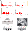
(A) ΔLc and F SMFS histograms of pFS-2 polyproteins carrying the Q/N-rich PLD of Orb2A show a broad mechanical polymorphism in terms of the increased contour length (ΔLc, top) and mechanical stability (bottom), ranging from NM conformers (orange bars) to different M conformers (red bars, n = 106), similar to that found in pathological amyloids [41]. In line with its reduced ability to form amyloids (see Fig 3 and S4 Fig), the mechanical conformational polymorphism of the F5Y mutant is diminished, increasing the proportion of NM conformers (n = 109). (B) Immunodot blot showed that like toxic oligomeric intermediates of other amyloidogenic proteins, both full-length Orb2A, and to a lesser extent Orb2B, as well as their isolated PLDs, are recognized by the A11 antibody [45]. (C) A representative electron micrograph of aged Orb2A PLD shows the formation oligomers (asterisks) and typical unbranched amyloid fibers (arrows) resembling those of the full-length Orb2A (see Fig 1G) and pathological amyloids. Scale bar: 0.2 μm. Like pathological amyloids, those species are recognized by the fiber-specific OC monoclonal antibody [46]. For panels B and C, Aβ42 oligomers and fibers were used as positive controls for the A11 and OC antibodies, respectively, while ubiquitin was used as a negative control. The underlying data for panels in this figure can be found in S1 Data.
Fig 5
Fig 5. Orb2 cytotoxic oligomeric species rapidly evolve into mature amyloids and they can be kinetically trapped as well as sequestered by pathological amyloids.
(A) Structural features during Orb2A assembly probed with conformational antibodies showed the formation of species reactive to A11 and OC antibodies. OC can exist in a SDS-resistant form, while A11-reactive oligomers are SDS-sensitive. (B) Lifespan of Aβ42 A11-reactive species. (C) Chemical structure of EGCG (left) and amphotericin B (AmB, right), used to trap OC- and A11-reactive oligomers, respectively. (D) Immunodot blot analysis of Orb2A:EGCG and Orb2A:AmB complexes probed with A11 and OC conformational antibodies show that oligomers trapped in the presence of EGCG interact with the OC antibody (and not with A11), whereas those formed in the presence of AmB react strongly with the A11 antibody (and not with OC antibody). (E) Survival curves of COS-7 cells microinjected with several samples (the A11-reactivity of which is indicated). Data are represented as the mean ± SEM: ***p < 0.001 (green asterisks, Orb2A:AmB versus remaining samples; Two-way ANOVA and Bonferroni post-test). Dextran, AmB, the intrinsically disordered region of Vamp2 (Vamp2Cyt) and dimethylsulfoxide (DMSO) are used as controls. The number of single-cells microinjected per sample was n = 100–200. (F) Fluorescence micrographs of COS-7 cells microinjected with different samples and fluorescein-labelled dextran (at 0 and 24 h after microinjection). Microinjection of the Orb2A:AmB complex resulted in a marked drop in the number of live cells at 24 h compared to those at 0 h. Notably, the A11 antibody rescued cells from apoptosis. Scale bars: 100 μm. (G) Huntingtin protein-containing expanded polyQ repeats (HttQ128) enhances Orb2A aggregation, and this enhancement requires the Orb2A PLD. Orb2A and HttQ128 coaggregates. HttQ does not induce aggregation of other EGFP-tagged neuronal proteins. Substitution of Orb2A PLD with the NM region of yeast Sup35 showed similar HttQ-dependent aggregation. Scale bar: large panel 20 μm, inset 10 μm. The underlying data for panels in this figure can be found in S1 Data.
Fig 6
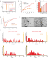
Fig 6. QBP1 interferes with Orb2 amyloid formation in vitro.
(A) Representative calorimetric traces for the interaction of full-length Orb2A with the QBP1 and SCR peptides. Traces in orange and pink correspond to the heat released upon injection of Orb2A into the ITC cell loaded with an excess of QBP1 or SCR, respectively, while black, blue, and red traces correspond to the Orb2A, QBP1, and SCR dilutions, respectively. (B) QBP1 but not SCR drastically reduced the turbidity at 405 nm of aged Orb2A PLD. (C) QBP1 reduces the quantity of amyloid formed as shown by measuring the CR bound to Orb2A PLD. The data are represented as the mean ± SEM: *p < 0.05 and ***p < 0.001 (One-way ANOVA and Tukey post-test). (D) Far-UV CD spectroscopy in the absence or presence of QBP1 or SCR indicates that QBP1 blocks protein precipitation over time of Orb2A PLD, but SCR does not. (E) Representative electron micrographs show that oligomers (asterisks) and amyloid fibers (arrows) of Orb2A PLD were drastically reduced when incubated with QBP1 (left) but not with the SCR (right). Scale bars: 1 μm. (F) The conformational polymorphism of Orb2A PLD is strongly diminished in the presence of a 1:10 molar excess of QBP1, which decreases the M frequency relative to the NM conformers (QBP1, n = 112 and SCR, n = 101). The underlying data for panels in this figure can be found in S1 Data.
Fig 7
Fig 7. QBP1 interferes with Orb2-mediated memory consolidation in vivo.
Pan-neuronal expression of QBP1 disrupts long-term male courtship suppression memory, while it does not interfere with short-term memory. Expression of QBP1 in the Δ80Q orb2 mutant background did not have any additive effect on long-term memory. The data are represented as the mean ± SEM. We assumed statistical significance at *p < 0.05 (One-way ANOVA). n = 16, 15, 25, 22, 18, 14, 15, 28, 27, 20 from left to right. The underlying data for panels in this figure can be found in S1 Data.
Fig 8
Fig 8. Structural basis of memory consolidation mediated by Orb2 amyloid.
In Drosophila, the Orb2 protein (top) is a functional amyloid with self-sustaining prion-like properties that follows an amyloidogenic pathway resembling that of pathological amyloids (bottom). During the assembly pathway, Orb2 and pathological amyloids can form A11-reactive toxic oligomers, and Orb2 can be sequestered by pathological amyloids to form hetero-oligomers. However, intrinsic structural features make Orb2 toxic oligomers rare and transient, suggesting that this functional amyloid appears to have been honed by evolution in adopting a self-sustaining amyloid-like state much more efficiently than pathological amyloids in order to avoid cytotoxicity and perform its role in memory consolidation. This function is supported by the amyloid state of Orb2, since its inhibition by an anti-amyloidogenic peptide (QBP1) selectively interferes with the long-term persistence of memory.

References

    1. Chernoff YO. Amyloidogenic domains, prions and structural inheritance: rudiments of early life or recent acquisition? Curr Opin Chem Biol. 2004;8(6):665–71. - PubMed
    1. Greenwald J, Riek R. On the possible amyloid origin of protein folds. J Mol Biol. 2012;421(4–5):417–26. 10.1016/j.jmb.2012.04.015 - DOI - PubMed
    1. Lansbury PT, Lashuel HA. A century-old debate on protein aggregation and neurodegeneration enters the clinic. Nature. 2006;443(7113):774–9. - PubMed
    1. Pepys MB. Amyloidosis. Annual review of medicine. 2006;57:223–41. - PubMed
    1. Fowler DM, Koulov AV, Balch WE, Kelly JW. Functional amyloid—from bacteria to humans. Trends Biochem Sci. 2007;32(5):217–24. - PubMed

Publication types

MeSH terms