

All Hormone-Producing Cell Types of the Pituitary Intermediate and Anterior Lobes Derive From Prop1-Expressing Progenitors
- PMID: 26812162
- PMCID: PMC4816735
- DOI: 10.1210/en.2015-1862
All Hormone-Producing Cell Types of the Pituitary Intermediate and Anterior Lobes Derive From Prop1-Expressing Progenitors
Abstract
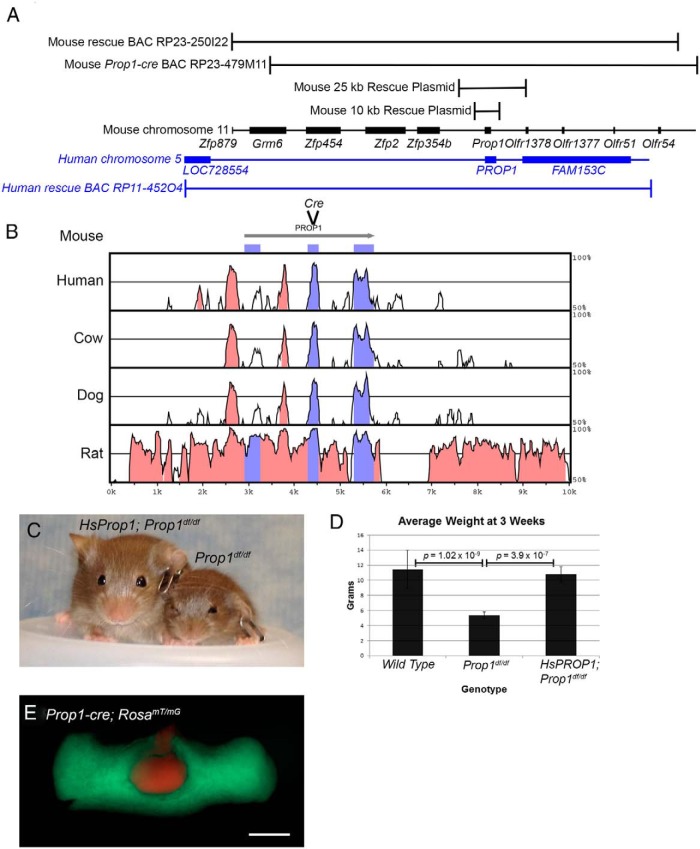
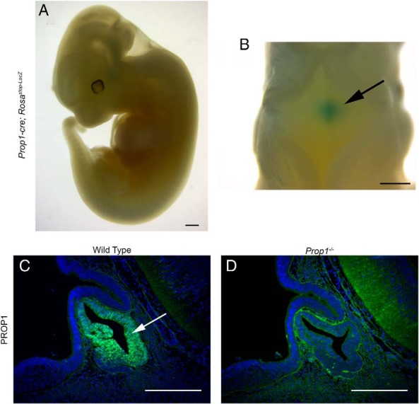
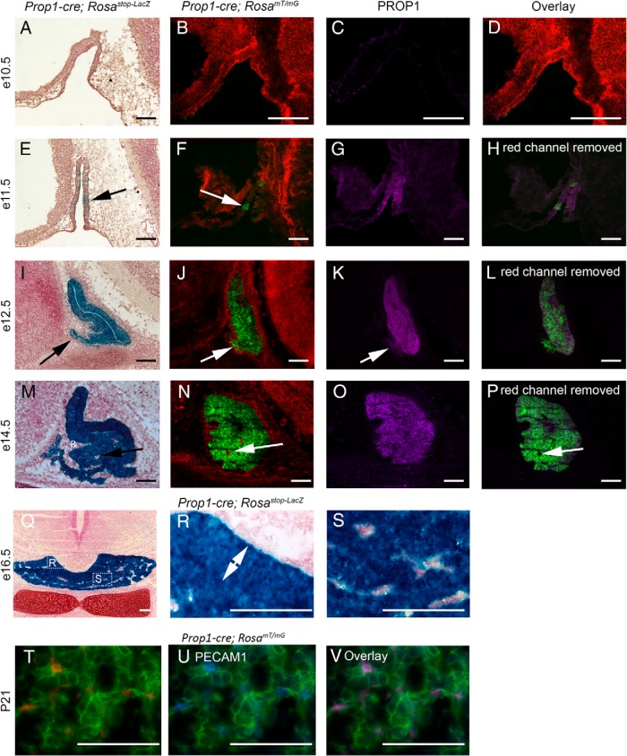
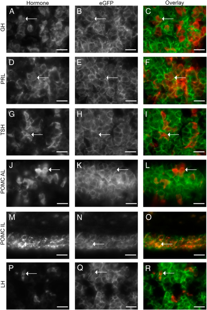
Mutations in PROP1, the most common known cause of combined pituitary hormone deficiency in humans, can result in the progressive loss of all hormones of the pituitary anterior lobe. In mice, Prop1 mutations result in the failure to initiate transcription of Pou1f1 (also known as Pit1) and lack somatotropins, lactotropins, and thyrotropins. The basis for this species difference is unknown. We hypothesized that Prop1 is expressed in a progenitor cell that can develop into all anterior lobe cell types, and not just the somatotropes, thyrotropes, and lactotropes, which are collectively known as the PIT1 lineage. To test this idea, we produced a transgenic Prop1-cre mouse line and conducted lineage-tracing experiments of Prop1-expressing cells. The results reveal that all hormone-secreting cell types of both the anterior and intermediate lobes are descended from Prop1-expressing progenitors. The Prop1-cre mice also provide a valuable genetic reagent with a unique spatial and temporal expression for generating tissue-specific gene rearrangements early in pituitary gland development. We also determined that the minimal essential sequences for reliable Prop1 expression lie within 10 kilobases of the mouse gene and demonstrated that human PROP1 can substitute functionally for mouse Prop1. These studies enhance our understanding of the pathophysiology of disease in patients with PROP1 mutations.
Figures
Similar articles
-
PROP1 coexists with SOX2 and induces PIT1-commitment cells.Biochem Biophys Res Commun. 2009 Jul 17;385(1):11-5. doi: 10.1016/j.bbrc.2009.05.027. Epub 2009 May 12. Biochem Biophys Res Commun. 2009. PMID: 19442651
-
Rapid transition of NESTIN-expressing dividing cells from PROP1-positive to PIT1-positive advances prenatal pituitary development.J Neuroendocrinol. 2013 Sep;25(9):779-91. doi: 10.1111/jne.12077. J Neuroendocrinol. 2013. PMID: 23855824
-
Role of PROP1 in Postnatal Pituitary Gland Maturation.Endocrinology. 2025 Jul 8;166(9):bqaf047. doi: 10.1210/endocr/bqaf047. Endocrinology. 2025. PMID: 40048699
-
Pituitary transcription factors in the aetiology of combined pituitary hormone deficiency.Best Pract Res Clin Endocrinol Metab. 2011 Feb;25(1):43-60. doi: 10.1016/j.beem.2010.10.014. Best Pract Res Clin Endocrinol Metab. 2011. PMID: 21396574 Review.
-
Regulation of pituitary stem cells by epithelial to mesenchymal transition events and signaling pathways.Mol Cell Endocrinol. 2017 Apr 15;445:14-26. doi: 10.1016/j.mce.2016.09.016. Epub 2016 Sep 17. Mol Cell Endocrinol. 2017. PMID: 27650955 Free PMC article. Review.
Cited by
-
Gut bacteria induce IgA expression in pituitary hormone-secreting cells during aging.iScience. 2023 Aug 26;26(10):107747. doi: 10.1016/j.isci.2023.107747. eCollection 2023 Oct 20. iScience. 2023. PMID: 37692284 Free PMC article.
-
The Musashi RNA binding proteins direct the translational activation of key pituitary mRNAs.Sci Rep. 2024 Mar 11;14(1):5918. doi: 10.1038/s41598-024-56002-8. Sci Rep. 2024. PMID: 38467682 Free PMC article.
-
Heterozygous variants in SIX3 and POU1F1 cause pituitary hormone deficiency in mouse and man.Hum Mol Genet. 2023 Jan 13;32(3):367-385. doi: 10.1093/hmg/ddac192. Hum Mol Genet. 2023. PMID: 35951005 Free PMC article.
-
Nucleoredoxin regulates WNT signaling during pituitary stem cell differentiation.bioRxiv [Preprint]. 2025 Feb 3:2025.01.30.635771. doi: 10.1101/2025.01.30.635771. bioRxiv. 2025. Update in: Hum Mol Genet. 2025 May 6;34(10):870-881. doi: 10.1093/hmg/ddaf032. PMID: 39975280 Free PMC article. Updated. Preprint.
-
Mouse models of growth hormone deficiency.Rev Endocr Metab Disord. 2021 Mar;22(1):3-16. doi: 10.1007/s11154-020-09601-5. Epub 2020 Oct 9. Rev Endocr Metab Disord. 2021. PMID: 33033978 Review.
References
-
- Alatzoglou KS, Dattani MT. Genetic forms of hypopituitarism and their manifestation in the neonatal period. Early Hum Dev. 2009;85:705–712. - PubMed
-
- Vallette-Kasic S, Brue T, Pulichino AM, et al. Congenital isolated adrenocorticotropin deficiency: an underestimated cause of neonatal death, explained by TPIT gene mutations. J Clin Endocrinol Metab. 2005;90:1323–1331. - PubMed
-
- Li S, Crenshaw EB, 3rd, Rawson EJ, Simmons DM, Swanson LW, Rosenfeld MG. Dwarf locus mutants lacking three pituitary cell types result from mutations in the POU-domain gene pit-1. Nature. 1990;347:528–533. - PubMed
-
- Pfäffle RW, DiMattia GE, Parks JS, et al. Mutation of the POU-specific domain of Pit-1 and hypopituitarism without pituitary hypoplasia. Science. 1992;257:1118–1121. - PubMed
-
- Sornson MW, Wu W, Dasen JS, et al. Pituitary lineage determination by the Prophet of Pit-1 homeodomain factor defective in Ames dwarfism. Nature. 1996;384:327–333. - PubMed
Publication types
MeSH terms
Substances
Grants and funding
LinkOut - more resources
Full Text Sources
Other Literature Sources
Medical
Molecular Biology Databases
