

Reference Charts for Fetal Cerebellar Vermis Height: A Prospective Cross-Sectional Study of 10605 Fetuses
- PMID: 26812238
- PMCID: PMC4727931
- DOI: 10.1371/journal.pone.0147528
Reference Charts for Fetal Cerebellar Vermis Height: A Prospective Cross-Sectional Study of 10605 Fetuses
Abstract
Objective: To establish reference charts for fetal cerebellar vermis height in an unselected population.
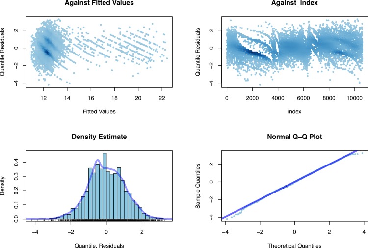
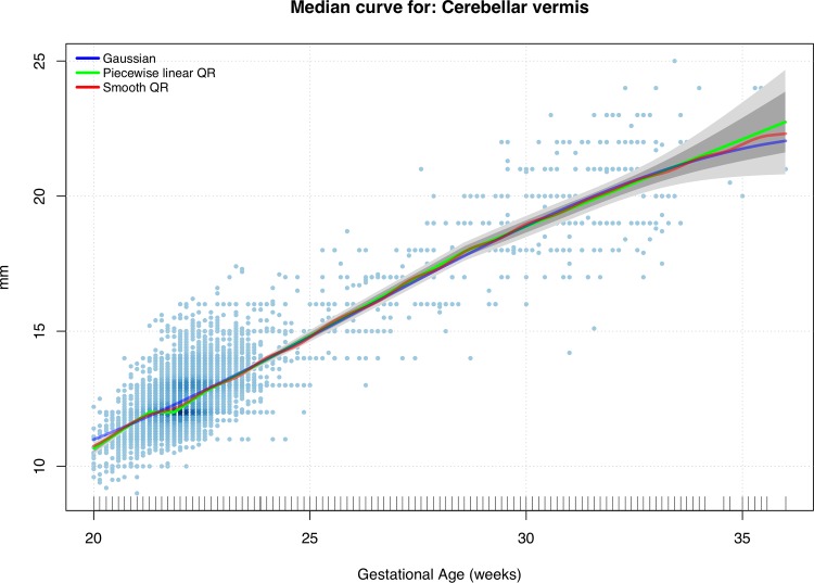
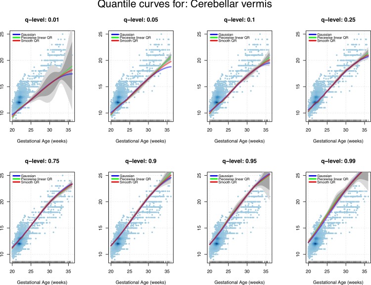
Methods: A prospective cross-sectional study between September 2009 and December 2014 was carried out at ALTAMEDICA Fetal-Maternal Medical Centre, Rome, Italy. Of 25203 fetal biometric measurements, 12167 (48%) measurements of the cerebellar vermis were available. After excluding 1562 (12.8%) measurements, a total of 10605 (87.2%) fetuses were considered and analyzed once only. Parametric and nonparametric quantile regression models were used for the statistical analysis. In order to evaluate the robustness of the proposed reference charts regarding various distributional assumptions on the ultrasound measurements at hand, we compared the gestational age-specific reference curves we produced through the statistical methods used. Normal mean height based on parametric and nonparametric methods were defined for each week of gestation and the regression equation expressing the height of the cerebellar vermis as a function of gestational age was calculated. Finally the correlation between dimension/gestation was measured.
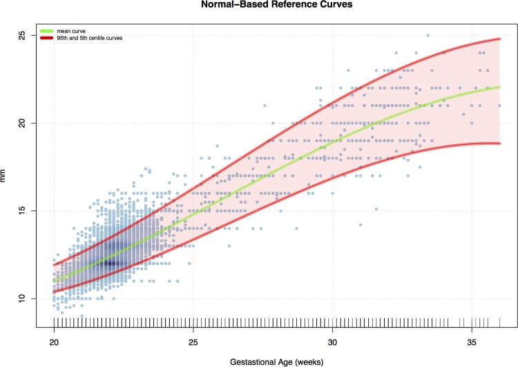
Results: The mean height of the cerebellar vermis was 12.7mm (SD, 1.6mm; 95% confidence interval, 12.7-12.8mm). The regression equation expressing the height of the CV as a function of the gestational age was: height (mm) = -4.85+0.78 x gestational age. The correlation between dimension/gestation was expressed by the coefficient r = 0.87.
Conclusion: This is the first prospective cross-sectional study on fetal cerebellar vermis biometry with such a large sample size reported in literature. It is a detailed statistical survey and contains new centile-based reference charts for fetal height of cerebellar vermis measurements.
Conflict of interest statement
Figures

Similar articles
-
Reference charts for fetal corpus callosum length: a prospective cross-sectional study of 2950 fetuses.J Ultrasound Med. 2014 Jun;33(6):1065-78. doi: 10.7863/ultra.33.6.1065. J Ultrasound Med. 2014. PMID: 24866614
-
The development of the fetal vermis: an in-utero sonographic evaluation.Ultrasound Obstet Gynecol. 2002 Feb;19(2):136-9. doi: 10.1046/j.0960-7692.2001.00621.x. Ultrasound Obstet Gynecol. 2002. PMID: 11876804
-
Development of the Fetal Vermis: New Biometry Reference Data and Comparison of 3 Diagnostic Modalities-3D Ultrasound, 2D Ultrasound, and MR Imaging.AJNR Am J Neuroradiol. 2016 Jul;37(7):1359-66. doi: 10.3174/ajnr.A4725. Epub 2016 Mar 31. AJNR Am J Neuroradiol. 2016. PMID: 27032974 Free PMC article.
-
Reference Ranges for Vermis Biometry on Prenatal Ultrasound: Systematic Review and Meta-Analysis.Ultraschall Med. 2023 Feb;44(1):e25-e38. doi: 10.1055/a-1408-1998. Epub 2021 Apr 9. Ultraschall Med. 2023. PMID: 33836547 English.
-
The World Health Organization fetal growth charts: concept, findings, interpretation, and application.Am J Obstet Gynecol. 2018 Feb;218(2S):S619-S629. doi: 10.1016/j.ajog.2017.12.010. Am J Obstet Gynecol. 2018. PMID: 29422204 Review.
Cited by
-
Mathematical models of human cerebellar development in the fetal period.J Anat. 2018 Apr;232(4):596-603. doi: 10.1111/joa.12767. Epub 2018 Jan 8. J Anat. 2018. PMID: 29315634 Free PMC article.
-
Quantitative fetal magnetic resonance imaging assessment of cystic posterior fossa malformations.Ultrasound Obstet Gynecol. 2020 Jul;56(1):78-85. doi: 10.1002/uog.21890. Ultrasound Obstet Gynecol. 2020. PMID: 31595598 Free PMC article.
-
Biometry of the Cerebellar Vermis and Brain Stem in Children: MR Imaging Reference Data from Measurements in 718 Children.AJNR Am J Neuroradiol. 2019 Nov;40(11):1835-1841. doi: 10.3174/ajnr.A6257. Epub 2019 Oct 17. AJNR Am J Neuroradiol. 2019. PMID: 31624120 Free PMC article.
-
Early alterations in cortical and cerebellar regional brain growth in Down Syndrome: An in vivo fetal and neonatal MRI assessment.Neuroimage Clin. 2020;25:102139. doi: 10.1016/j.nicl.2019.102139. Epub 2019 Dec 23. Neuroimage Clin. 2020. PMID: 31887718 Free PMC article.
-
The volume of the cerebellum in the second semester of gestation.Clujul Med. 2018;91(2):176-180. doi: 10.15386/cjmed-922. Epub 2018 Apr 25. Clujul Med. 2018. PMID: 29785155 Free PMC article.
References
-
- Sgaier SK, Millet S, Villanueva MP, Berenshteyn F, Song C, Joyner AL. Morphogenetic and cellular movements that shape the mouse cerebellum; insights from genetic fate mapping. Neuron. 2005. January 6;45(1):27–40. - PubMed
-
- Barkovic. Pediatric Neuroimaging 3rd Ed. Philadelphia: Lippincott Williams & Wilkins; 2000; 337–41.
-
- American Institute of Ultrasound in Medicine. AIUM practice guidelines for the performance of an antepartum obstetric ultrasound examination J Ultrasound Med 2010;29:157–66. - PubMed
-
- Rapoport M, van Reekum R, Mayberg H. The role of the cerebellum in cognition and behavior: a selective review. J Neuropsychiatry ClinNeurosci. 2000;12(2):193–8. - PubMed
MeSH terms
LinkOut - more resources
Full Text Sources
Other Literature Sources
