

Cog-Wheel Octameric Structure of RS1, the Discoidin Domain Containing Retinal Protein Associated with X-Linked Retinoschisis
- PMID: 26812435
- PMCID: PMC4728063
- DOI: 10.1371/journal.pone.0147653
Cog-Wheel Octameric Structure of RS1, the Discoidin Domain Containing Retinal Protein Associated with X-Linked Retinoschisis
Abstract
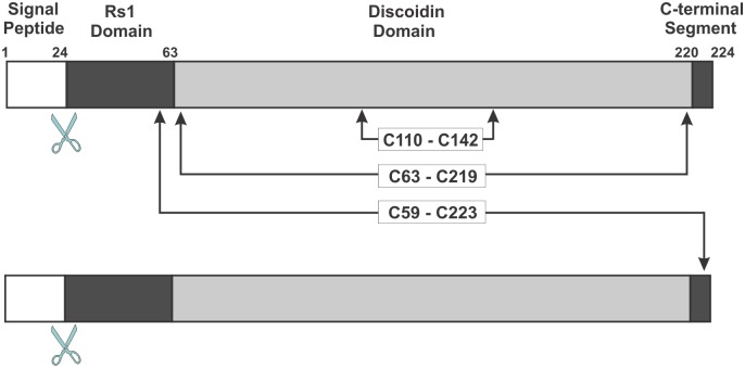
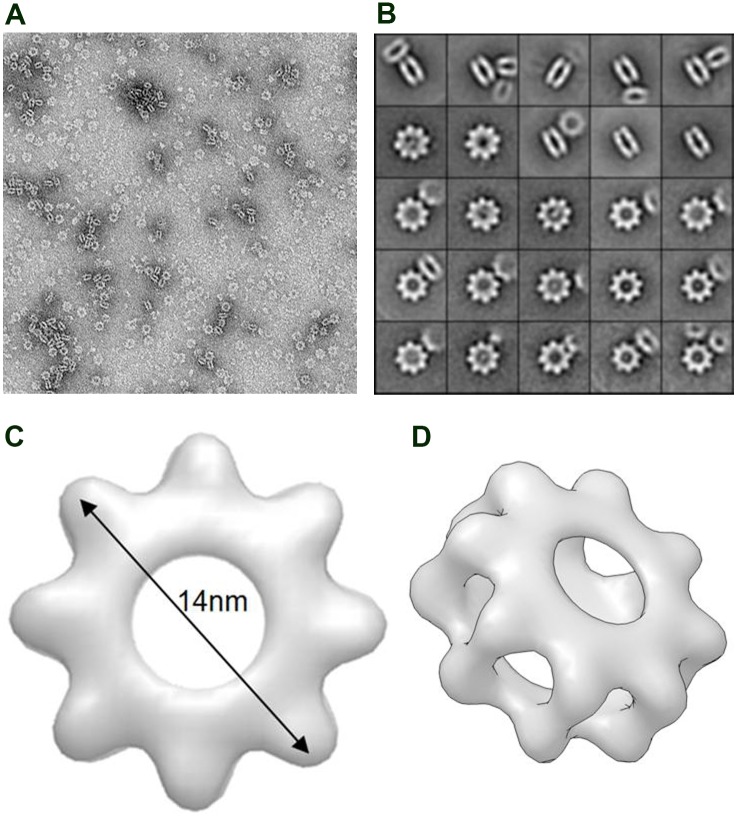
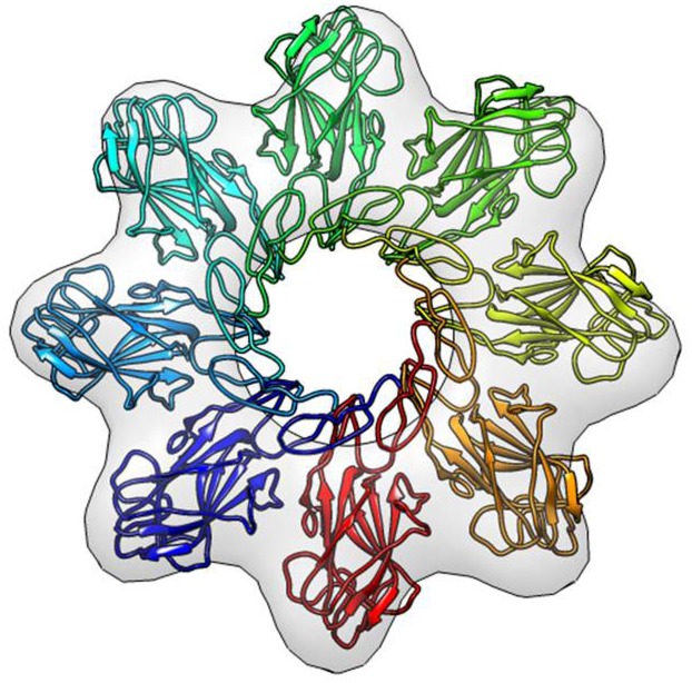
RS1, also known as retinoschisin, is a disulphide-linked, discoidin domain containing homo-oligomeric protein that plays a crucial role in maintaining the cellular and synaptic organization of the retina. This is highlighted by the finding that over 130 mutations in RS1 cause X-linked retinoschisis, a retinal degenerative disease characterized by the splitting of the retinal cell layers, disruption of the photoreceptor-bipolar synapses, degeneration of photoreceptors, and severe loss in central vision. In this study, we investigated the arrangement of the RS1 subunits within the oligomer complex using single particle electron microscopy. RS1 was seen as two stacked rings with each ring displaying a symmetrical cog wheel-like structure with eight teeth or projections corresponding to the RS1 subunits. Three dimensional reconstruction and molecular modelling indicated that the discoidin domain, the principal functional unit of RS1, projects outward, and the Rs1 domain and C-terminal segment containing intermolecular disulphide bonds are present in the inner ring to form the core octameric structure. These studies provide a basis for further understanding the role of the novel core RS1 octameric complex in retinal cell biology and X-linked retinoschisis.
Conflict of interest statement
Figures
Similar articles
-
RS1, a discoidin domain-containing retinal cell adhesion protein associated with X-linked retinoschisis, exists as a novel disulfide-linked octamer.J Biol Chem. 2005 Mar 18;280(11):10721-30. doi: 10.1074/jbc.M413117200. Epub 2005 Jan 11. J Biol Chem. 2005. PMID: 15644328
-
Defective discoidin domain structure, subunit assembly, and endoplasmic reticulum processing of retinoschisin are primary mechanisms responsible for X-linked retinoschisis.J Biol Chem. 2003 Jul 25;278(30):28139-46. doi: 10.1074/jbc.M302464200. Epub 2003 May 13. J Biol Chem. 2003. PMID: 12746437
-
X-linked juvenile retinoschisis: clinical diagnosis, genetic analysis, and molecular mechanisms.Prog Retin Eye Res. 2012 May;31(3):195-212. doi: 10.1016/j.preteyeres.2011.12.002. Epub 2012 Jan 3. Prog Retin Eye Res. 2012. PMID: 22245536 Free PMC article. Review.
-
Retinoschisin (RS1), the protein encoded by the X-linked retinoschisis gene, is anchored to the surface of retinal photoreceptor and bipolar cells through its interactions with a Na/K ATPase-SARM1 complex.J Biol Chem. 2007 Nov 9;282(45):32792-801. doi: 10.1074/jbc.M706321200. Epub 2007 Sep 5. J Biol Chem. 2007. PMID: 17804407
-
Advances in understanding the molecular structure of retinoschisin while questions remain of biological function.Prog Retin Eye Res. 2023 Jul;95:101147. doi: 10.1016/j.preteyeres.2022.101147. Epub 2022 Nov 16. Prog Retin Eye Res. 2023. PMID: 36402656 Free PMC article. Review.
Cited by
-
Targeted Expression of Retinoschisin by Retinal Bipolar Cells in XLRS Promotes Resolution of Retinoschisis Cysts Sans RS1 From Photoreceptors.Invest Ophthalmol Vis Sci. 2022 Oct 3;63(11):8. doi: 10.1167/iovs.63.11.8. Invest Ophthalmol Vis Sci. 2022. PMID: 36227606 Free PMC article.
-
Structural analysis of X-linked retinoschisis mutations reveals distinct classes which differentially effect retinoschisin function.Hum Mol Genet. 2016 Dec 15;25(24):5311-5320. doi: 10.1093/hmg/ddw345. Hum Mol Genet. 2016. PMID: 27798099 Free PMC article.
-
Retinoschisin Facilitates the Function of L-Type Voltage-Gated Calcium Channels.Front Cell Neurosci. 2017 Aug 8;11:232. doi: 10.3389/fncel.2017.00232. eCollection 2017. Front Cell Neurosci. 2017. PMID: 28848397 Free PMC article.
-
Predominant Founder Effect among Recurrent Pathogenic Variants for an X-Linked Disorder.Genes (Basel). 2022 Apr 12;13(4):675. doi: 10.3390/genes13040675. Genes (Basel). 2022. PMID: 35456481 Free PMC article.
-
Multimodal imaging in a pedigree of X-linked Retinoschisis with a novel RS1 variant.BMC Med Genet. 2018 Nov 12;19(1):195. doi: 10.1186/s12881-018-0712-8. BMC Med Genet. 2018. PMID: 30419843 Free PMC article.
References
-
- Zeng Y, Takada Y, Kjellstrom S, Hiriyanna K, Tanikawa A, Wawrousek E, et al. RS-1 Gene Delivery to an Adult Rs1h Knockout Mouse Model Restores ERG b-Wave with Reversal of the Electronegative Waveform of X-Linked Retinoschisis. Invest Ophthalmol Vis Sci. 2004;45(9):3279–85. doi: 10.1167/iovs.04-0576 45/9/3279 [pii]. . - DOI - PubMed
-
- Weber BH, Schrewe H, Molday LL, Gehrig A, White KL, Seeliger MW, et al. Inactivation of the murine X-linked juvenile retinoschisis gene, Rs1h, suggests a role of retinoschisin in retinal cell layer organization and synaptic structure. Proc Natl Acad Sci U S A. 2002;99(9):6222–7. 10.1073/pnas.092528599 99/9/6222 [pii]. . - DOI - PMC - PubMed
-
- Kellner U, Brummer S, Foerster MH, Wessing A. X-linked congenital retinoschisis. Graefes Arch Clin Exp Ophthalmol. 1990;228(5):432–7. Epub 1990/01/01. . - PubMed
Publication types
MeSH terms
Substances
Grants and funding
LinkOut - more resources
Full Text Sources
Other Literature Sources
Molecular Biology Databases
