

Toward FRP-Based Brain-Machine Interfaces-Single-Trial Classification of Fixation-Related Potentials
- PMID: 26812487
- PMCID: PMC4727887
- DOI: 10.1371/journal.pone.0146848
Toward FRP-Based Brain-Machine Interfaces-Single-Trial Classification of Fixation-Related Potentials
Abstract
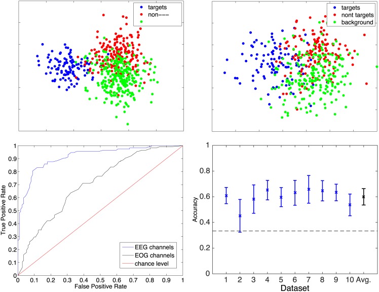
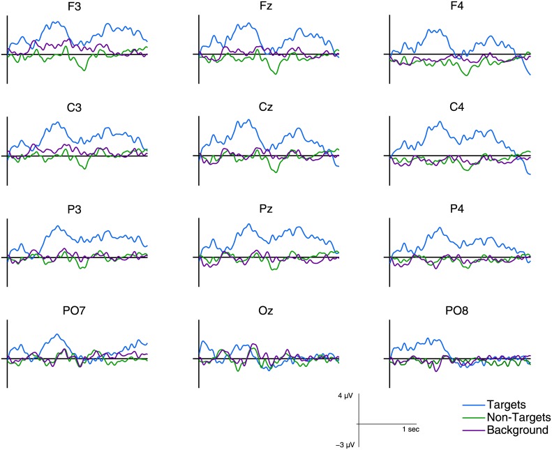
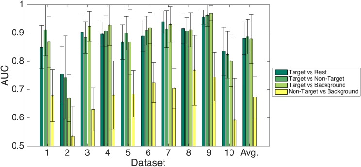
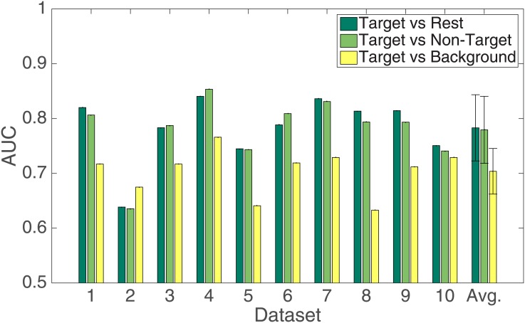
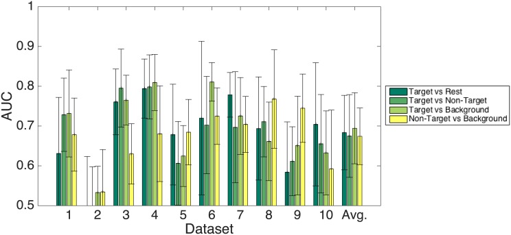
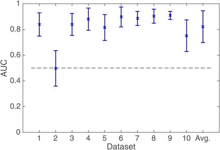
The co-registration of eye tracking and electroencephalography provides a holistic measure of ongoing cognitive processes. Recently, fixation-related potentials have been introduced to quantify the neural activity in such bi-modal recordings. Fixation-related potentials are time-locked to fixation onsets, just like event-related potentials are locked to stimulus onsets. Compared to existing electroencephalography-based brain-machine interfaces that depend on visual stimuli, fixation-related potentials have the advantages that they can be used in free, unconstrained viewing conditions and can also be classified on a single-trial level. Thus, fixation-related potentials have the potential to allow for conceptually different brain-machine interfaces that directly interpret cortical activity related to the visual processing of specific objects. However, existing research has investigated fixation-related potentials only with very restricted and highly unnatural stimuli in simple search tasks while participant's body movements were restricted. We present a study where we relieved many of these restrictions while retaining some control by using a gaze-contingent visual search task. In our study, participants had to find a target object out of 12 complex and everyday objects presented on a screen while the electrical activity of the brain and eye movements were recorded simultaneously. Our results show that our proposed method for the classification of fixation-related potentials can clearly discriminate between fixations on relevant, non-relevant and background areas. Furthermore, we show that our classification approach generalizes not only to different test sets from the same participant, but also across participants. These results promise to open novel avenues for exploiting fixation-related potentials in electroencephalography-based brain-machine interfaces and thus providing a novel means for intuitive human-machine interaction.
Conflict of interest statement
Figures

Similar articles
-
EEG Negativity in Fixations Used for Gaze-Based Control: Toward Converting Intentions into Actions with an Eye-Brain-Computer Interface.Front Neurosci. 2016 Nov 18;10:528. doi: 10.3389/fnins.2016.00528. eCollection 2016. Front Neurosci. 2016. PMID: 27917105 Free PMC article.
-
An eye fixation-related potentials analysis of the P300 potential for fixations onto a target object when exploring natural scenes.J Vis. 2015;15(13):20. doi: 10.1167/15.13.20. J Vis. 2015. PMID: 26401627
-
Face Selective Neural Activity: Comparisons Between Fixed and Free Viewing.Brain Topogr. 2020 May;33(3):336-354. doi: 10.1007/s10548-020-00764-7. Epub 2020 Mar 31. Brain Topogr. 2020. PMID: 32236786
-
Coregistration of eye movements and EEG in natural reading: analyses and review.J Exp Psychol Gen. 2011 Nov;140(4):552-72. doi: 10.1037/a0023885. J Exp Psychol Gen. 2011. PMID: 21744985 Review.
-
Eye Movements and Fixation-Related Potentials in Reading: A Review.Vision (Basel). 2020 Feb 3;4(1):11. doi: 10.3390/vision4010011. Vision (Basel). 2020. PMID: 32028566 Free PMC article. Review.
Cited by
-
EEG Negativity in Fixations Used for Gaze-Based Control: Toward Converting Intentions into Actions with an Eye-Brain-Computer Interface.Front Neurosci. 2016 Nov 18;10:528. doi: 10.3389/fnins.2016.00528. eCollection 2016. Front Neurosci. 2016. PMID: 27917105 Free PMC article.
-
Using Fixation-Related Potentials for Inspecting Natural Interactions.Front Hum Neurosci. 2020 Nov 5;14:579505. doi: 10.3389/fnhum.2020.579505. eCollection 2020. Front Hum Neurosci. 2020. PMID: 33250729 Free PMC article.
-
Co-registration of eye movements and neuroimaging for studying contextual predictions in natural reading.Lang Cogn Neurosci. 2019 May 16;35(5):595-612. doi: 10.1080/23273798.2019.1616102. eCollection 2020. Lang Cogn Neurosci. 2019. PMID: 32656295 Free PMC article.
-
An error-aware gaze-based keyboard by means of a hybrid BCI system.Sci Rep. 2018 Sep 4;8(1):13176. doi: 10.1038/s41598-018-31425-2. Sci Rep. 2018. PMID: 30181532 Free PMC article.
-
Is Neural Activity Detected by ERP-Based Brain-Computer Interfaces Task Specific?PLoS One. 2016 Oct 28;11(10):e0165556. doi: 10.1371/journal.pone.0165556. eCollection 2016. PLoS One. 2016. PMID: 27792781 Free PMC article.
References
-
- Mueller-Putz GR, Scherer R, Brauneis C, Pfurtscheller G. Steady-state visual evoked potential (SSVEP)-based communication: impact of harmonic frequency components. Journal of Neural Engineering. 2005;2(4):123 Available from: http://stacks.iop.org/1741-2552/2/i=4/a=008. 10.1088/1741-2560/2/4/008 - DOI - PubMed
-
- Hwanga HJ, Lima JH, Junga YJ, Choia H, Leeb SW, Im CH. Development of an SSVEP-based BCI spelling system adopting a QWERTY-style LED keyboard. Journal of Neuroscience Methods. 2012;208:59–65. - PubMed
Publication types
MeSH terms
LinkOut - more resources
Full Text Sources
Other Literature Sources
Miscellaneous
