. 2016 Apr 1:129:367-377.
doi: 10.1016/j.neuroimage.2016.01.020. Epub 2016 Jan 23.

Sleep reverts changes in human gray and white matter caused by wake-dependent training

Affiliations

Sleep reverts changes in human gray and white matter caused by wake-dependent training

Giulio Bernardi et al. Neuroimage. .

Abstract

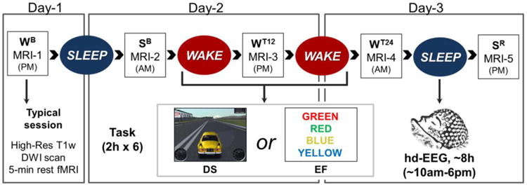
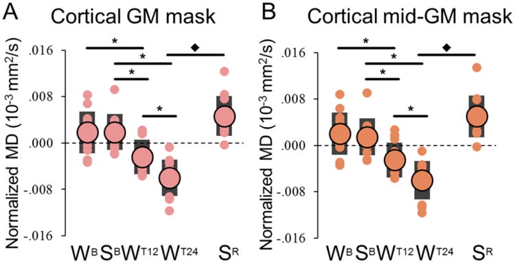
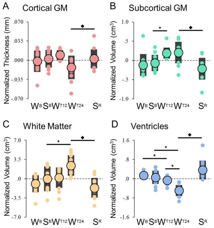
Learning leads to rapid microstructural changes in gray (GM) and white (WM) matter. Do these changes continue to accumulate if task training continues, and can they be reverted by sleep? We addressed these questions by combining structural and diffusion weighted MRI and high-density EEG in 16 subjects studied during the physiological sleep/wake cycle, after 12 h and 24 h of intense practice in two different tasks, and after post-training sleep. Compared to baseline wake, 12 h of training led to a decline in cortical mean diffusivity. The decrease became even more significant after 24 h of task practice combined with sleep deprivation. Prolonged practice also resulted in decreased ventricular volume and increased GM and WM subcortical volumes. All changes reverted after recovery sleep. Moreover, these structural alterations predicted cognitive performance at the individual level, suggesting that sleep's ability to counteract performance deficits is linked to its effects on the brain microstructure. The cellular mechanisms that account for the structural effects of sleep are unknown, but they may be linked to its role in promoting the production of cerebrospinal fluid and the decrease in synapse size and strength, as well as to its recently discovered ability to enhance the extracellular space and the clearance of brain metabolites.

Keywords: DWI; Extracellular space; MRI; Mean diffusivity; Sleep deprivation.


Conflict of interest statement

Conflicts of interest. G. Tononi is involved in a research study in humans supported by Philips Respironics. This study is not related to the work presented in the current manuscript. R. Benca has served as a consultant to Merck and Jazz, and receives research funding from Merck. A. Alexander is part owner of inseRT MRI, Inc. This company is not related to the work and did not sponsor this research. The other authors have indicated no financial conflicts of interest.

Figures

Figure 1
Figure 1. Experimental design
Each subject completed two experiments (DS and EF), spaced at least 2 weeks apart. During both experiments MRI sessions were performed every ∼12 hours: WB (wake baseline), after a wake day spent outside the lab without any specific training; SB (sleep baseline) the next morning, after subjects slept at home as usual; WT12 (wake with training), after 12h of wake with extensive training in the lab; WT24 (extended wake with training), after 24h of continuous wake with extensive training in the lab; SR (sleep recovery), after ∼8h of recovery sleep with hd-EEG recording in the lab. Each MRI session included a high-resolution anatomical scan, a 5-min eyes-closed resting state functional scan, and a diffusion weighted scan (DWI).
Figure 2
Figure 2. Large-scale ROIs used for the analyses of global brain changes
The four ROIs (here shown in a representative subject) are: [green] subcortical grey matter (GM), [yellow] white matter (WM), [blue] ventricles and [red] cortical GM. Moreover, a mid-GM cortical mask was generated in order to eliminate voxels characterized by a high probability of containing mixed tissues (see text for details).
Figure 3
Figure 3. Comparison of MD variations detected using the cortical GM mask (A) and the mid-GM mask (B) excluding voxels along the WM-GM and GM-CSF interfaces
On average the mid-GM mask included 60.4 ± 1.4 % less voxels than the original cortical mask. Large circles represent the group-level average, while each small circle represents a different subject (black bars indicate one standard deviation from the mean). Values were zero-mean normalized by subtracting the across-time-points mean. *, significant differences for planned comparisons (p < 0.05, after Bonferroni-Holm adjustment; N = 12); ◆, significant effect of recovery sleep (paired t-test).
Figure 4
Figure 4. Global changes in cortical thickness (A) and volume changes in subcortical GM, WM and ventricles (B-D)
Large circles represent the group-level average, while each small circle represents a different subject (black bars indicate one standard deviation). Values were zero-mean normalized by subtracting the across-time-points mean. *, significant differences for planned comparisons (p < 0.05, after Bonferroni-Holm adjustment; N = 12); ◆, significant effect of recovery sleep (paired t-test).
Figure 5
Figure 5. Prediction of vigilance level based on structural parameters
For both DS (top) and EF (bottom), the graphs show the “real” (purple) vigilance level observed during sessions SB, WT12, WT24 and SR (measured as the reaction time during the PVT test), in comparison with the vigilance level predicted based on global structural parameters (green), in a representative subject (S06).

References

    1. Alexander AL, Hasan KM, Lazar M, Tsuruda JS, Parker DL. Analysis of partial volume effects in diffusion-tensor MRI. Magn Reson Med. 2001;45:770–780. - PubMed
    1. Anderson BJ, Li X, Alcantara AA, Isaacs KR, Black JE, Greenough WT. Glial hypertrophy is associated with synaptogenesis following motor-skill learning, but not with angiogenesis following exercise. Glia. 1994;11:73–80. - PubMed
    1. Assaf Y, Pasternak O. Diffusion tensor imaging (DTI)-based white matter mapping in brain research: a review. J Mol Neurosci. 2008;34:51–61. - PubMed
    1. Ball G, Srinivasan L, Aljabar P, Counsell SJ, Durighel G, Hajnal JV, Rutherford MA, Edwards AD. Development of cortical microstructure in the preterm human brain. Proceedings of the National Academy of Sciences. 2013;110:9541–9546. - PMC - PubMed
    1. Basner M, Dinges DF. Maximizing Sensitivity of the Psychomotor Vigilance Test (PVT) to Sleep Loss. Sleep. 2011;34:581–591. - PMC - PubMed

Publication types