. 2016 Jan 27:6:19781.
doi: 10.1038/srep19781.

Skeletal muscle Heat shock protein 60 increases after endurance training and induces peroxisome proliferator-activated receptor gamma coactivator 1 α1 expression

Affiliations

Skeletal muscle Heat shock protein 60 increases after endurance training and induces peroxisome proliferator-activated receptor gamma coactivator 1 α1 expression

Rosario Barone et al. Sci Rep. .

Abstract

Heat shock protein 60 (Hsp60) is a chaperone localizing in skeletal muscle mitochondria, whose role is poorly understood. In the present study, the levels of Hsp60 in fibres of the entire posterior group of hindlimb muscles (gastrocnemius, soleus, and plantaris) were evaluated in mice after completing a 6-week endurance training program. The correlation between Hsp60 levels and the expression of four isoforms of peroxisome proliferator-activated receptor gamma coactivator 1 alpha (PGC1α) were investigated only in soleus. Short-term overexpression of hsp60, achieved by in vitro plasmid transfection, was then performed to determine whether this chaperone could have a role in the activation of the expression levels of PGC1α isoforms. The levels of Hsp60 protein were fibre-type specific in the posterior muscles and endurance training increased its content in type I muscle fibers. Concomitantly with the increased levels of Hsp60 released in the blood stream of trained mice, mitochondrial copy number and the expression of three isoforms of PGC1α increased. Overexpressing hsp60 in cultured myoblasts induced only the expression of PGC1 1α, suggesting a correlation between Hsp60 overexpression and PGC1 1 α activation.


Figures
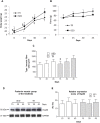
Figure 1
Figure 1. Functional effects of endurance exercise on the body weight, strength, cross-sectional area (CSA) and Hsp60 protein levels in the posterior group of hindlimb muscles.
(A), changes in the body weight over time. Square (▄) sedentary (SED) mice; Rhombus (♦) trained (TR) mice; horizontal axis, time of training. Data are presented as the means ± SD. # significantly different from SED15 (n = 8), SED30 (n = 8) and SED45 (n = 8) mice (P < 0.001); * significantly different than TR30 (n = 8) and TR45 (n = 8) mice (P < 0.001); § significantly different from SED30 and SED45 mice (P < 0.001); ‡ significantly different from TR45 mice (P < 0.001); † significantly different from SED45 mice (P < 0.05). (B) strength of the forelimbs. Square (▄) SED mice; Rhombus (♦) TR mice; horizontal axis, time of training. Data are presented as the means ± SD. ◊ significantly different from TR15 (n = 8), TR30 and TR45 (P < 0.001). (C) histogram showing the results for the CSA of type I fibers of the posterior group of hindlimb muscles. An average of 435 fibers was analyzed for each mouse. Bars, groups of mice; open bars, SED mice; shaded bars, TR mice; horizontal axis, time of training. (D) representative western blots of Hsp60 (60 kDa) levels in the posterior group of hindlimb muscles (gastrocnemius, soleus, and plantaris) at various time points (n = 3). 60 μg of protein was loaded in each lane; GAPDH (37 kDa) was used as the loading control. (E) relative expression levels of Hsp60 from the sedentary and trained groups of mice at various times of training, as shown in the horizontal axis. AU: Arbitrary Unit.
Figure 2
Figure 2. Immunohistochemistry and densitometric analysis of the staining intensity in the posterior group of hindlimb muscles demonstrate that Hsp60 is elevated in type IIa and I muscle fibers, and with endurance training.
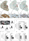
(A) immunohistochemistry for Hsp60 (I), MHC-I (II) and MHC-IIa/x (III) in serial cross-sections of the posterior group of hindlimb muscles, reconstructed by combining multiple images captured at low magnification (10×) (gastrocnemius, soleus, and plantaris, are circled in green, red, and blue, respectively). (B) enhanced magnification of the images shown in A. Type IIb fibers are negative to antibodies anti-MHC-I and anti-MHC-IIa/x. Bar 25 μm. (C) Representative images used for densitometric analysis of the staining intensity of a cross-section immunostained for Hsp60. The analysis was performed using 5 fields per section, 5 sections per mice (40 μm between sections) and 8 mice per group. An average of 435 fibers was analyzed for each mouse. The acquired RGB image (Bb) was transformed to a greyscale image (32-bit) and then inverted. Representative histograms of the fibers I, IIa, IIb, and IIx are shown in the small framed panels, where 0 corresponds to no positivity and 255 corresponds to maximal positivity. (D), the staining intensity (determined with ImageJ 1.41 software) of the fibers was expressed as the mean pixel intensity (PI) normalized to the cross-sectional area (CSA) for the red gastrocnemius (I), the plantaris (II), and the soleus (III). SED45 and TR45 indicate trained and sedentary mice on day 45, respectively. Data are presented as the means ± SD. #significantly different from type I fibers from SED45 mice (P < 0.01), *(P < 0.05), **(P < 0.001).
Figure 3
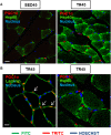
Figure 3. Confocal microscopy analysis further demonstrates that Hsp60 protein levels increase in mice trained for 45 days and shows the localization of Hsp60 in the myofibrillar cell.
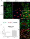
(A) immunofluorescence for Hsp60 and MHC-I of cross-sections of sedentary and trained mice at 45 days (SED45, n = 8, and TR45, n = 8, respectively); the arrows indicate the type I fibers. Bar 25 μm. (B) the staining intensity for Hsp60 (bars) of type I fibers was expressed as the mean pixel intensity (PI) normalized to the CSA (cross-sectional area) using the software Leica application suite advanced fluorescences software. Open bar, sedentary (SED45) mice; shaded bar, trained (TR45) mice; both on day 45. Data are presented as the means ± SD. *significantly different from SED45 mice (P < 0.05). (C) representative immunofluorescence image revealing that Hsp60 was localized also in inter-myofibrillar mitochondria (IMF). Bar 10 μm. (D) representative immunofluorescence image revealing that Hsp60 was localized to the subsarcolemmal (SS) space. Bar 20 μm.
Figure 4
Figure 4. Hsp60 protein levels increase in parallel with training intensity in the soleus muscle and in the blood stream of trained mice.
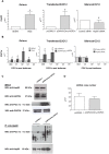
(A) representative western blots of Hsp60 (60 kDa) in the soleus from the trained (n = 8) and sedentary mice (n = 8) at various time points. 40 μg of protein was loaded in each lane; GAPDH (37 kDa) was used as the loading control. (B) relative levels of Hsp60 in the soleus. Open bars, sedentary mice; shaded bars, trained mice; horizontal axis, days. AU: Arbitrary Unit. (C) copy number of mitochondrial genes in the soleus of sedentary mice (n = 6) at day 45 (SED45, open bar) and trained mice (n = 6) at day 45 (TR45, shaded bar). (D) serum levels of Hsp60 in SED (n = 8) and TR (n = 8) groups at various time points. Open bars, sedentary (SED) mice; shaded bars, trained (TR) mice; horizontal axis, days. Data are presented as the means ± SD. ∂ significantly different from TR30 mice (P < 0.05). # significantly different from TR45 mice (P < 0.001).
Figure 5
Figure 5. qRT-PCR analysis validates the increase in the levels of hsp60 gene expression, shows the increase in the levels of PGC1α gene expression and its isoforms in the soleus of trained mice, and suggests a possible correlation between hsp60 and PGC1 α1 genes in HSPD1 transfected C2C12 cells.
(A) bars show the hsp60 gene expression levels normalized for the reference genes, according to the Livak Method (2−∆∆CT) (Schmittgen and Livak, 2008) in: soleus of sedentary (n = 6) and trained (n = 6) mice at 45 days (SED45, and TR45, respectively); C2C12 myoblasts transfected with pCMV6-Entry-HSPD1 vector to over-express hsp60 (pcDNA3.1 was used as a negative control); and Hsp60 siRNA for silencing Hsp60 (scramble siRNA used as a negative control, Control siRNA). (B) bars show the PGC1α isoforms [PGC1α total (αtot), isoform 1 (α1), 2 (α2), 3 (α3), 4 (α4)] gene expression normalized for the reference genes, according to the Livak Method (2−∆∆CT), in: soleus of SED45 (grey bars) and TR45 (black bars); C2C12 myoblast transfected with pCMV6-Entry-HSPD1 vector to over-express hsp60 (pcDNA3.1 was used as a negative control); and Hsp60 siRNA for silencing Hsp60 (scramble siRNA used as a negative control, Control siRNA). Data are presented as means ± SD, bars show the PGC1α isoforms [PGC1α total (P < 0.01); ∂ significantly different from SED45 (P < 0.01); #significantly different from pcDNA3.1 (P < 0.01); ¥significantly different from Control siRNA (P < 0.01). (C) Upper panel: western blot analysis for endogenous expression levels of both Hsp60 and PGC1α in whole-cell lysates from C2C12 myoblasts transfected with pCMV6-Entry-HSPD1 vector to over-express hsp60 (pcDNA3.1 was used as a negative control). GAPDH was used as a control for loading. Lower panel: representative blot of immunoprecipitation experiments in C2C12 myoblasts transfected with pCMV6-Entry-HSPD1 vector to over-express hsp60 (pcDNA3.1 was used as a negative control) showing a 113 kDa band corresponding to PGC1α that co-immunoprecipited with Hsp60. D, copy number of mitochondrial genes in C2C12 myoblast transfected with pCMV6-Entry-HSPD1 vector (shaded bar) (pcDNA3.1 was used as a negative control, open bar).
Figure 6
Figure 6. PGC1 α1 levels increase in the soleus in trained mice and in transfected C2C12 cells upon transfection with pCMV-Entry-HSPD1 vector.
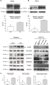
(A) representative western blots of soleus and relative expression levels (bars) of PGC1 α1 (113 kDa), 4 HNE (55 kDa), Mn SOD (25 kDa), p-AMPKα (63 kDa), AMPKα1 (63 kDa), AMPKα2 (63 kDa), TFAM (30 kDa) in soleus of sedentary (SED45, open bar, n = 8) and trained (TR45, shaded bar, n = 8) mice at 45 days. 80 μg of proteins were loaded in each lane; GAPDH (37 kDa) was used as the loading control. Data are presented as the means ± SD. ∂ significantly different from TR45 mice (P < 0.001). AU: Arbitrary Unit. (B) representative western blots of C2C12 cells and relative levels (bars) of PGC1 α1 (113 kDa), 4 HNE (55 kDa), Mn SOD (25 kDa), p-AMPKα (63 kDa), AMPKα1 (63 kDa), AMPKα2 (63 kDa), TFAM (30 kDa) in C2C12 myoblast transfected with pCMV6-Entry-HSPD1 vector (pcDNA3.1 was used as a negative control). 80 μg of proteins were loaded in each lane; GAPDH (37 kDa) was used as the loading control and TR45 was used as positive control. Data are presented as the means ± SD. #significantly different from pCMV6-Entry-HSPD1 (P < 0.0001). AU: Arbitrary Unit. *significant results. (C) relative expression levels of 4 HNE, p-AMPKα, p-AMPKα/(AMPKα1 + AMPKα2) in the soleus. Open bars, sedentary mice; shaded bars, trained mice. AU: Arbitrary Unit.
Figure 7
Figure 7. The confocal microscopy analysis shows the localization of PGC1α positive cells in the skeletal muscle tissue.
(A) immunofluorescence for Hsp60 and PGC1α of muscle cross-sections of sedentary and trained mice at 45 days (SED45 and TR45, respectively). Bar 25 μm. (B) immunofluorescence for laminin and PGC1α (the arrows indicate the localization of PGC1α in the interstitial space, outside the fibers; a negative control is shown in supplemental Figure S1B); and Hsp60 and PGC1α of muscle cross-sections of trained mice at 45 days (TR45) at higher magnification. Bar 15 μm.
Figure 8
Figure 8. Exosomal preparations from the blood of trained mice and qRT-PCR analysis of PGC1 α1 gene in C2C12 cells treated with the conditioned media from transfected C2C12 myoblasts.
(A) Transmission electron microscopy (TEM) image showing the morphology of exosomes purified from blood. (B) western blot analysis demonstrating the presence of Hsp60 (60 kDa) and of exosomal markers Alix (90 kDa) and Hsp70 (70 kDa) in the purified exosomes obtained from the blood of twenty-four (four mice for each blood sample) mice submitted to a single bout of exercise and sacrificed immediately (0′) and 15 min (15′) after completing exercise, and sedentary mice (CN). 80 μg of protein was loaded in each lane. Relative expression levels of Alix, Hsp70 and Hsp60 in exosomes derived from blood of trained and sedentary mice. AU: Arbitrary Unit. # significantly different from 0′ and 15′ (P < 0.001). * significantly different from 15′ (P < 0.001).

References

    1. Cuervo A. M. & Wong E. Chaperone-mediated autophagy: roles in disease and aging. Cell research 24, 92–104, 10.1038/cr.2013.153 (2014). - DOI - PMC - PubMed
    1. Czarnecka A. M., Campanella C., Zummo G. & Cappello F. Mitochondrial chaperones in cancer: from molecular biology to clinical diagnostics. Cancer biology & therapy 5, 714–720 (2006). - PubMed
    1. Georgopoulos C. & Welch W. J. Role of the major heat shock proteins as molecular chaperones. Annual review of cell biology 9, 601–634, 10.1146/annurev.cb.09.110193.003125 (1993). - DOI - PubMed
    1. Halperin L., Jung J. & Michalak M. The many functions of the endoplasmic reticulum chaperones and folding enzymes. IUBMB life 66, 318–326, 10.1002/iub.1272 (2014). - DOI - PubMed
    1. Kakkar V., Meister-Broekema M., Minoia M., Carra S. & Kampinga H. H. Barcoding heat shock proteins to human diseases: looking beyond the heat shock response. Disease models & mechanisms 7, 421–434, 10.1242/dmm.014563 (2014). - DOI - PMC - PubMed

Publication types

Substances