

Contribution of writing to reading: Dissociation between cognitive and motor process in the left dorsal premotor cortex
- PMID: 26813381
- PMCID: PMC6867475
- DOI: 10.1002/hbm.23118
Contribution of writing to reading: Dissociation between cognitive and motor process in the left dorsal premotor cortex
Abstract
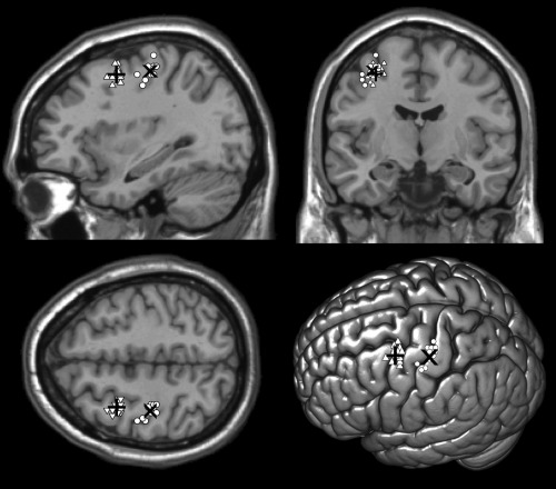
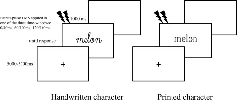
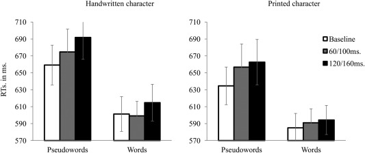
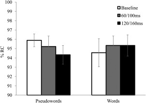
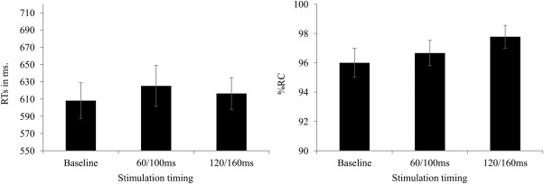
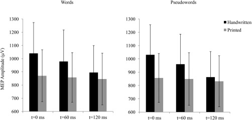
Functional brain imaging studies reported activation of the left dorsal premotor cortex (PMd), that is, a main area in the writing network, in reading tasks. However, it remains unclear whether this area is causally relevant for written stimulus recognition or its activation simply results from a passive coactivation of reading and writing networks. Here, we used chronometric paired-pulse transcranial magnetic stimulation (TMS) to address this issue by disrupting the activity of the PMd, the so-called Exner's area, while participants performed a lexical decision task. Both words and pseudowords were presented in printed and handwritten characters. The latter was assumed to be closely associated with motor representations of handwriting gestures. We found that TMS over the PMd in relatively early time-windows, i.e., between 60 and 160 ms after the stimulus onset, increased reaction times to pseudoword without affecting word recognition. Interestingly, this result pattern was found for both printed and handwritten characters, that is, regardless of whether the characters evoked motor representations of writing actions. Our result showed that under some circumstances the activation of the PMd does not simply result from passive association between reading and writing networks but has a functional role in the reading process. At least, at an early stage of written stimuli recognition, this role seems to depend on a common sublexical and serial process underlying writing and pseudoword reading rather than on an implicit evocation of writing actions during reading as typically assumed.
Keywords: Exner's area; cortico-spinal excitability; functional role; sublexical process; transcranial magnetic stimulation.
© 2016 Wiley Periodicals, Inc.
Figures
References
-
- Amunts K, Weiss PH, Mohlberg H, Pieperhoff P, Eickhoff S, Gurd JM, Marshall JC, Shah NJ, Fink GR, Zilles K (2004): Analysis of neural mechanisms underlying verbal fluency in cytoarchitectonically defined stereotaxic space‐The roles of Brodmann areas 44 and 45. Neuroimage 22:42–56. - PubMed
-
- Anderson SW, Damasio AR, Damasio H (1990): Troubled letters but not numbers: Domain specific cognitive impairments following focal damage in frontal cortex. Brain 113:749–766. - PubMed
-
- Balota DA, Cortese MJ, Sergent‐Marshall SD, Spieler DH, Yap M (2004): Visual word recognition of single‐syllable words. J Exp Psychol Gen 133:283–316. - PubMed
-
- Beeson P, Rapcsak S, Plante E, Chargualaf J, Chung A, Johnson S, Trouard T (2003): The neural substrates of writing: A functional magnetic resonance imaging study. Aphasiology 17:647–665.
Publication types
MeSH terms
LinkOut - more resources
Full Text Sources
Other Literature Sources
