. 2016 Jan 27:6:19814.
doi: 10.1038/srep19814.

Regulation of human cerebro-microvascular endothelial baso-lateral adhesion and barrier function by S1P through dual involvement of S1P1 and S1P2 receptors

Affiliations

Regulation of human cerebro-microvascular endothelial baso-lateral adhesion and barrier function by S1P through dual involvement of S1P1 and S1P2 receptors

Rachael Wiltshire et al. Sci Rep. .

Abstract

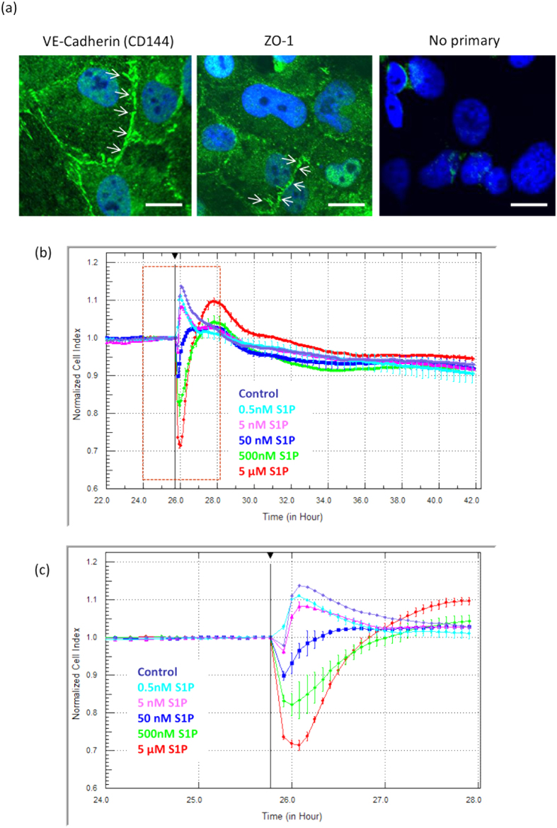
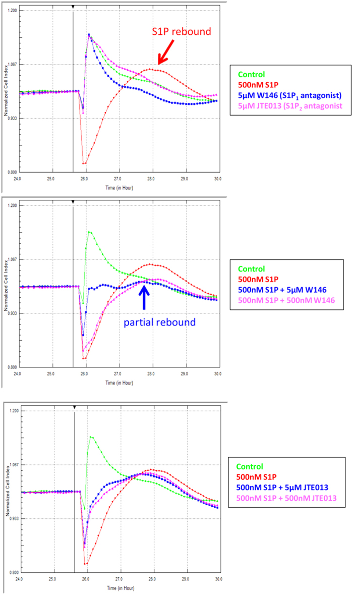
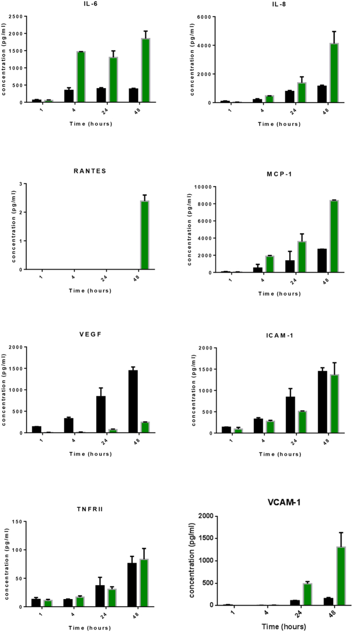
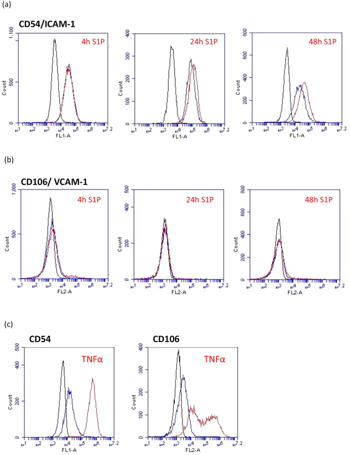
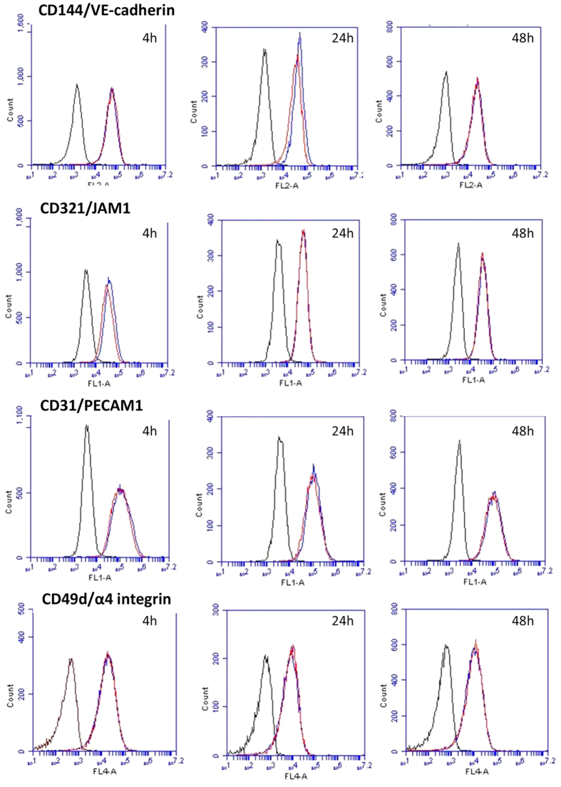
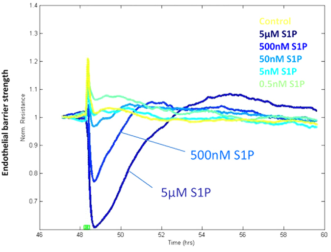
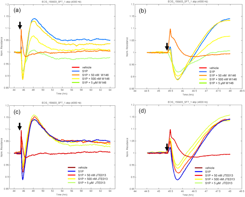
Herein we show that S1P rapidly and acutely reduces the focal adhesion strength and barrier tightness of brain endothelial cells. xCELLigence biosensor technology was used to measure focal adhesion, which was reduced by S1P acutely and this response was mediated through both S1P1 and S1P2 receptors. S1P increased secretion of several pro-inflammatory mediators from brain endothelial cells. However, the magnitude of this response was small in comparison to that mediated by TNFα or IL-1β. Furthermore, S1P did not significantly increase cell-surface expression of any key cell adhesion molecules involved in leukocyte recruitment, included ICAM-1 and VCAM-1. Finally, we reveal that S1P acutely and dynamically regulates microvascular endothelial barrier tightness in a manner consistent with regulated rapid opening followed by closing and strengthening of the barrier. We hypothesise that the role of the S1P receptors in this process is not to cause barrier dysfunction, but is related to controlled opening of the endothelial junctions. This was revealed using real-time measurement of barrier integrity using ECIS ZΘ TEER technology and endothelial viability using xCELLigence technology. Finally, we show that these responses do not occur simply though the pharmacology of a single S1P receptor but involves coordinated action of S1P1 and S1P2 receptors.


Figures

Figure 1
Figure 1. S1P rapidly and acutely alters brain endothelial focal adhesion.
The hCMVEC monolayers form tight and adherens junctions that are evident 24 hours after seeding. (a) shows the expression of the junctional proteins VE cadherin (CD144) and Zonula Occludin-1 (ZO-1) 24 hours after seeding of cells to demonstrate formation of junctional complexes. The no primary control stain is also shown for comparison. (b) S1P affects the focal adhesion of hCMVECs in a concentration and time-dependent manner. S1P was added at the time demarcated by the black line. (b) reveals the gross effect of S1P on hCMVEC adhesion during the 16h period following S1P addition. (c) reveals the rapid response to S1P during the first few hours after addition. These data are representative of 6 independent observations. Note these data are normalised to the Cell Index at the time of S1P addition. See supplemental figure 1 online for the actual Cell Index values (not normalised) and full time course data.
Figure 2
Figure 2. The reduction in focal adhesion induced by S1P requires both S1P1 and S1P2 receptors.
Receptor specific antagonists were used to ascertain the involvement of each respective receptor in the S1P (500 nM) response. (a) There were no effects of the antagonists (5 μM) on basal focal adhesion. (b) The S1P1 antagonist (W146) partially blocked the effect of S1P. (c) The S1P2 antagonist (JTE013) also partially blocked the effect of S1P. Antagonists for S1P3 and S1P4 receptors had no effect on the S1P response (data not shown). These responses have been observed in 4 independent experiments.
Figure 3
Figure 3. S1P increases secretion of specific inflammatory mediators.
Secreted cytokines and soluble surface adhesion molecules were measured using multiplex cytometric bead array technology. Conditioned media was collected 1h, 4 h, 24 h and 48 hours after treatment with 500 nM S1P (green bars). The secretions from the vehicle treated cells (0.005% DMSO) are the black bars. Data show the mean ± SEM from 3 independent experiments.
Figure 4
Figure 4. S1P regulation of key leukocyte tethering/adhesion molecules.
The surface expression of (a) ICAM-1 and (b) VCAM-1 were measured by flow cytometry 4 h, 24 h and 48 h after treatment with 500 nM S1P. Panels in (c) show the pronounced effect of the potent pro-inflammatory mediator TNFα on CD54/ICAM-1 and CD106/VCAM-1 expression by the hCMVEC as a comparison. In each histogram the black population is the background cellular auto fluorescence, the blue curve is the basal ICAM-1 or VCAM-1 expression and the red curve is the expression following treatment with S1P (a + b) or TNFα (c). These responses have been observed in 3 independent experiments.
Figure 5
Figure 5. S1P has no gross effect on the expression level of other key cell surface endothelial adhesion molecules.
Cell surface expression of CD144, CD321, CD31, CD49d were measured 4 h, 24 h and 48 hours after treatment with 500 nM S1P. Each cell adhesion molecule is expressed by the hCMVECs. In each histogram the black population is the background cellular auto fluorescence, the blue curve is the basal expression (CD144/CD321/CD31/CD49d) and the red curve is the expression following treatment with S1P (500 nM). These responses have been observed in 3 independent experiments.
Figure 6
Figure 6. S1P rapidly and acutely reduces the trans-endothelial electrical resistance, which is followed by strengthening of the endothelial barrier.
The temporal ECIS data shows rapid and acute reduction in endothelial barrier resistance following S1P addition (dotted lines) at 500 nM and 5 μM. This is followed by slower strengthening of the barrier resistance (3–4 hours) resulting in a stronger barrier resistance than before S1P treatment. Data show mean ± SD (4 wells). These responses have been observed in 6 independent ECIS experiments.
Figure 7
Figure 7. S1P induced changes in barrier function mediated through both S1P1 and S1P2 receptors.
The barrier strength of the hCMVECs measured using ECIS. Normalised TEER of 1 is equivalent to ~900Ω. (a,b) influence of the S1P1 antagonist W146 (50 nM to 5 μM) on the S1P response. (c,d) influence of the S1P2 antagonist JTE 013 (50 nM to 5 μM) on the S1P response. The (b) and (d) panels focus on the 3 h period following drug addition, to highlight the acute effects of the antagonists. Data are normalised to the time of drug addition. Similar responses were obtained in 3 independent experiments.

References

    1. Brinkmann V. sphingosine 1-phosphate receptors in health and disease. Mechanistic insights from gene deletion studies and reverse pharmacology. pharmacology and therapeutics 115, 84–105 (2007). - PubMed
    1. Fernández-Pisonero I. et al. Lipopolysaccharide and Sphingosine-1-Phosphate Cooperate To Induce Inflammatory Molecules and Leukocyte Adhesion in Endothelial Cells. The Journal of Immunology 189, 5402–5410, 10.4049/jimmunol.1201309 (2012). - DOI - PubMed
    1. Maceyka M., H. K., Milstien S. & Spiegel S. Sphingosine-1-phosphate signaling and its role in disease. trends cell biol 22, 10.1016/j.tcb.2011.09.003. (2012). - DOI - PMC - PubMed
    1. Murata N., Sato K., Kon J., Tomura H. & Okajima F. Quantitative Measurement of Sphingosine 1-Phosphate by Radioreceptor-Binding Assay. Analytical Biochemistry 282, 115–120, 10.1006/abio.2000.4580 (2000). - DOI - PubMed
    1. Murata N. et al. Interaction of sphingosine 1-phosphate with plasma components, including lipoproteins, regulates the lipid receptor-mediated actions. Biochem. J. 352, 809–815 (2000). - PMC - PubMed

Publication types