

Neisseria cinerea isolates can adhere to human epithelial cells by type IV pilus-independent mechanisms
- PMID: 26813911
- PMCID: PMC4891991
- DOI: 10.1099/mic.0.000248
Neisseria cinerea isolates can adhere to human epithelial cells by type IV pilus-independent mechanisms
Abstract
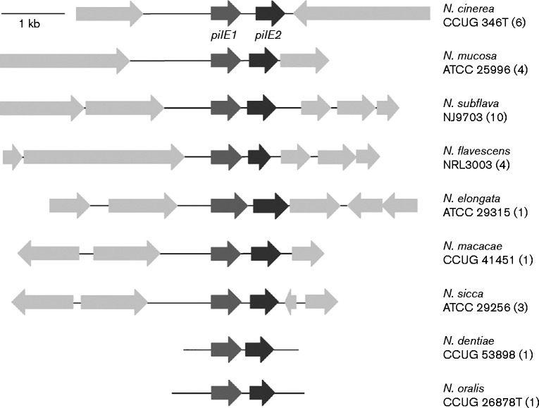
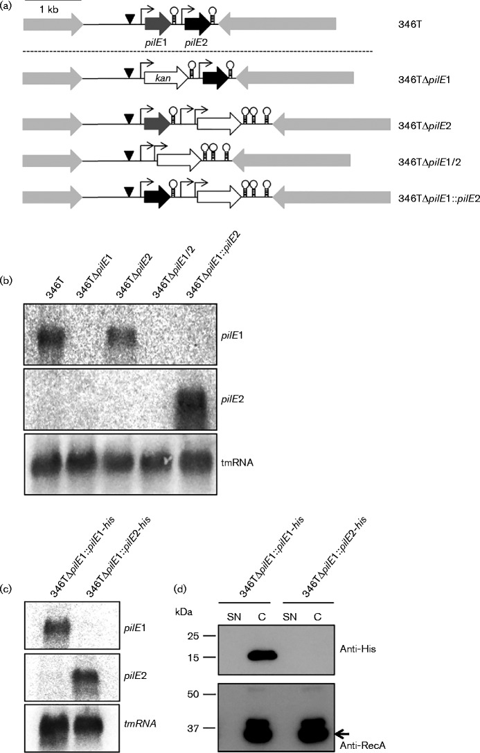
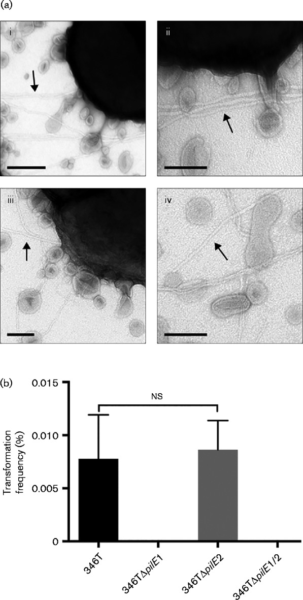
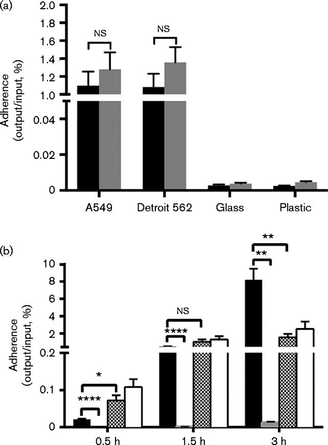
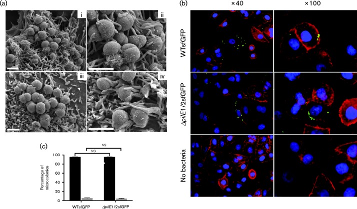
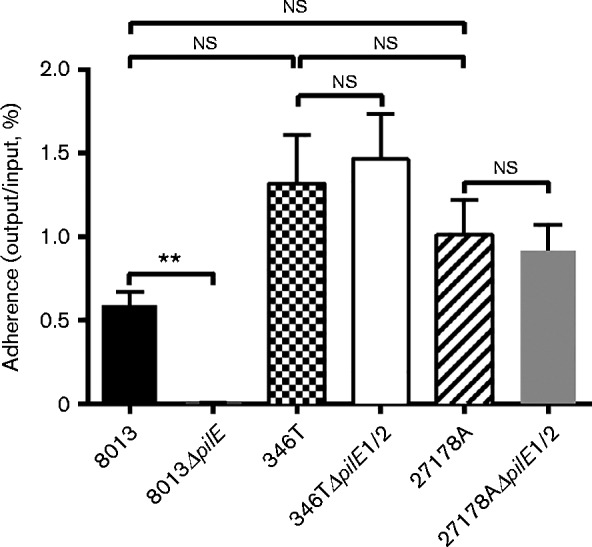
In pathogenic Neisseria species the type IV pili (Tfp) are of primary importance in host-pathogen interactions. Tfp mediate initial bacterial attachment to cell surfaces and formation of microcolonies via pilus-pilus interactions. Based on genome analysis, many non-pathogenic Neisseria species are predicted to express Tfp, but aside from studies on Neisseria elongata, relatively little is known about the formation and function of pili in these organisms. Here, we have analysed pilin expression and the role of Tfp in Neisseria cinerea. This non-pathogenic species shares a close taxonomic relationship to the pathogen Neisseria meningitidis and also colonizes the human oropharyngeal cavity. Through analysis of non-pathogenic Neisseria genomes we identified two genes with homology to pilE, which encodes the major pilin of N. meningitidis. We show which of the two genes is required for Tfp expression in N. cinerea and that Tfp in this species are required for DNA competence, similar to other Neisseria. However, in contrast to the meningococcus, deletion of the pilin gene did not impact the association of N. cinerea to human epithelial cells, demonstrating that N. cinerea isolates can adhere to human epithelial cells by Tfp-independent mechanisms.
Figures
Similar articles
-
Commensal Neisseria cinerea impairs Neisseria meningitidis microcolony development and reduces pathogen colonisation of epithelial cells.PLoS Pathog. 2020 Mar 24;16(3):e1008372. doi: 10.1371/journal.ppat.1008372. eCollection 2020 Mar. PLoS Pathog. 2020. PMID: 32208456 Free PMC article.
-
Contribution of σ70 and σN Factors to Expression of Class II pilE in Neisseria meningitidis.J Bacteriol. 2019 Sep 20;201(20):e00170-19. doi: 10.1128/JB.00170-19. Print 2019 Oct 15. J Bacteriol. 2019. PMID: 31331980 Free PMC article.
-
The hypervariable region of meningococcal major pilin PilE controls the host cell response via antigenic variation.mBio. 2014 Feb 11;5(1):e01024-13. doi: 10.1128/mBio.01024-13. mBio. 2014. PMID: 24520062 Free PMC article.
-
The role of pilin glycan in neisserial pathogenesis.Mol Cell Biochem. 2003 Nov;253(1-2):179-90. doi: 10.1023/a:1026058311857. Mol Cell Biochem. 2003. PMID: 14619968 Review.
-
Type-4 pili and meningococcal adhesiveness.Gene. 1997 Jun 11;192(1):149-53. doi: 10.1016/s0378-1119(96)00802-5. Gene. 1997. PMID: 9224885 Review.
Cited by
-
Rolling the evolutionary dice: Neisseria commensals as proxies for elucidating the underpinnings of antibiotic resistance mechanisms and evolution in human pathogens.Microbiol Spectr. 2024 Feb 6;12(2):e0350723. doi: 10.1128/spectrum.03507-23. Epub 2024 Jan 5. Microbiol Spectr. 2024. PMID: 38179941 Free PMC article.
-
Virulence genes and previously unexplored gene clusters in four commensal Neisseria spp. isolated from the human throat expand the neisserial gene repertoire.Microb Genom. 2020 Sep;6(9):mgen000423. doi: 10.1099/mgen.0.000423. Epub 2020 Aug 26. Microb Genom. 2020. PMID: 32845827 Free PMC article.
-
Genetic Manipulation of Neisseria gonorrhoeae and Commensal Neisseria Species.Curr Protoc. 2024 Sep;4(9):e70000. doi: 10.1002/cpz1.70000. Curr Protoc. 2024. PMID: 39228292 Free PMC article.
-
RpoN and the Nps and Npa two-component regulatory system control pilE transcription in commensal Neisseria.Microbiologyopen. 2019 May;8(5):e00713. doi: 10.1002/mbo3.713. Epub 2018 Aug 5. Microbiologyopen. 2019. PMID: 30079633 Free PMC article.
-
Neisseria cinerea Expresses a Functional Factor H Binding Protein Which Is Recognized by Immune Responses Elicited by Meningococcal Vaccines.Infect Immun. 2017 Sep 20;85(10):e00305-17. doi: 10.1128/IAI.00305-17. Print 2017 Oct. Infect Immun. 2017. PMID: 28739825 Free PMC article.
References
Publication types
MeSH terms
Substances
Grants and funding
LinkOut - more resources
Full Text Sources
Other Literature Sources
Molecular Biology Databases
Research Materials