. 2016 Jan 27:6:19722.
doi: 10.1038/srep19722.

Proteome-wide analysis reveals widespread lysine acetylation of major protein complexes in the malaria parasite

Affiliations

Proteome-wide analysis reveals widespread lysine acetylation of major protein complexes in the malaria parasite

Simon A Cobbold et al. Sci Rep. .

Abstract

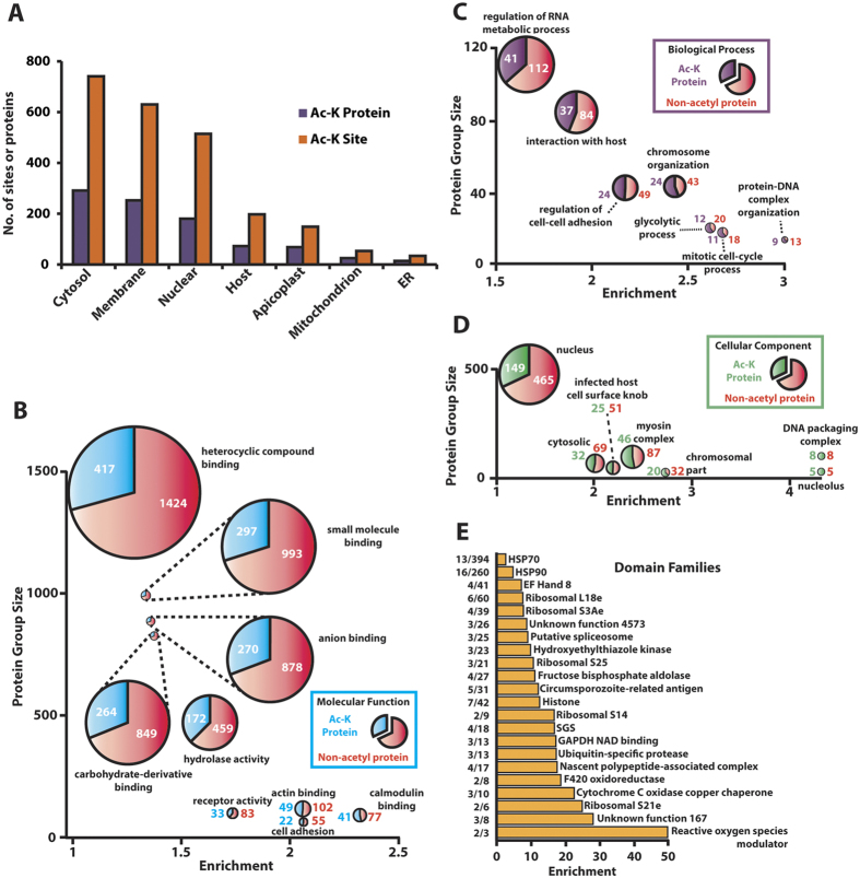
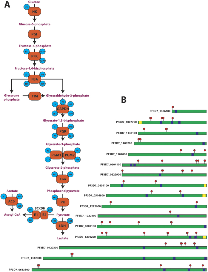
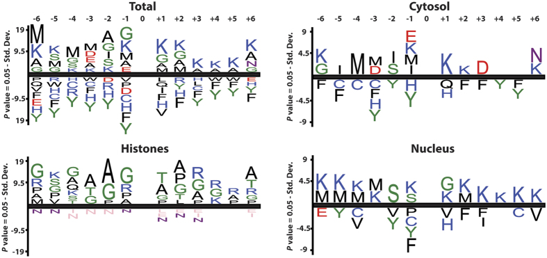
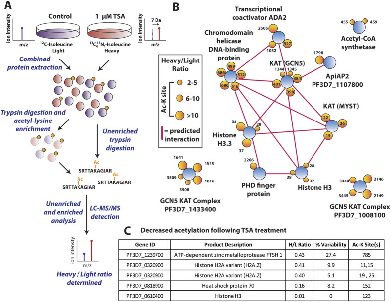
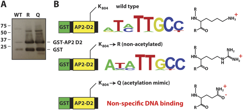
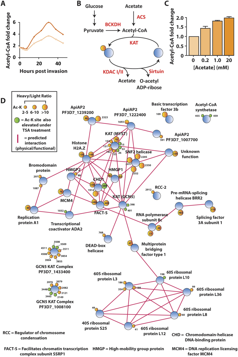
Lysine acetylation is a ubiquitous post-translational modification in many organisms including the malaria parasite Plasmodium falciparum, yet the full extent of acetylation across the parasite proteome remains unresolved. Moreover, the functional significance of acetylation or how specific acetyl-lysine sites are regulated is largely unknown. Here we report a seven-fold expansion of the known parasite 'acetylome', characterizing 2,876 acetylation sites on 1,146 proteins. We observe that lysine acetylation targets a diverse range of protein complexes and is particularly enriched within the Apicomplexan AP2 (ApiAP2) DNA-binding protein family. Using quantitative proteomics we determined that artificial perturbation of the acetate/acetyl-CoA balance alters the acetyl-lysine occupancy of several ApiAP2 DNA-binding proteins and related transcriptional proteins. This metabolic signaling could mediate significant downstream transcriptional responses, as we show that acetylation of an ApiAP2 DNA-binding domain ablates its DNA-binding propensity. Lastly, we investigated the acetyl-lysine targets of each class of lysine deacetylase in order to begin to explore how each class of enzyme contributes to regulating the P. falciparum acetylome.


Figures

Figure 1
Figure 1
(A) Distribution of acetylated protein and acetyl-lysine sites across subcellular compartments. Predicted localization of acetylated proteins from annotated GO terms. Enrichment of (B) molecular function (C) biological process (D) cellular component GO terms for acetylated proteins. Statistically significant enrichment of 22 terms across the three GO term groups was determined using hypergeometric testing (q < 0.01). 5 redundant groups were removed for clarity. (E) Acetyl-lysine enrichment of protein domains was assessed using three different approaches (see methods). Presented are the protein domains significantly enriched for acetylated lysines using the tiered stratified q-value method. Pfam families are listed as significantly enriched if their E-value is <1. The y-axis represents a ratio of the lysines present in each domain family acetylated to the total number of lysines within the domain family.
Figure 2
Figure 2
(A) Acetylation of glycolytic and acetyl-CoA biosynthetic enzymes. Numbers inside blue circles indicate the position of the acetyl-lysine site, enzymes represented are hexokinase (HK), phosphoglucose isomerase (PGI), phosphofructokinase (PFK), fructose bisphosphate aldolase (FBA), triose phosphate isomerase (TIM), glyceraldehyde-3-phosphate dehydrogenase (GAPDH), phosphoglycerate kinase (PGK), phosphoglycerate mutase 1/2 (PGM1/2), enolase (Eno), pyruvate kinase 1 (PK), lactate dehydrogenase (LDH), branched-chain keto-acid dehydrogenase subunit E1/E2 (BCKDH E1/E2), acetyl-CoA synthetase (ACS). (B) Distribution of acetyl-lysine on ApiAP2 DNA-binding proteins. Red flags indicate acetylated lysines, blue boxes represent AP2 DNA-binding domains and yellow boxes represent ACDC domains (AP2-coincident domain mostly at the C-terminus). The non-represented ApiAP2 proteins are not acetylated.
Figure 3
Figure 3. Sequence motifs surrounding acetylated lysines.
Sequence motif surrounding lysine acetylation presented as significantly enriched/de-enriched amino acids at positions −6 through +6 as percentage difference (p < 0.05).
Figure 4
Figure 4
(A) Stable isotope labeling by amino acids in cell culture. P. falciparum infected erythrocytes are maintained under standard culturing conditions in RPMI containing either 12C614N1 isoleucine (light) or 13C615N1 isoleucine (heavy) for 72 hours. After proteome labeling (>95%), the light culture was untreated and the heavy culture was treated with 1 μM of tricostatin A (TSA) for three hours. Treated (heavy) and untreated (light) cultures were saponin-permeabilized, washed, combined and extracted. Following trypsin digestion and SCX fractionation, samples were analyzed both directly (unenriched) and subsequent to enrichment by anti-acetyl lysine immunoprecipitation via high resolution nano-UPLC-MS and MS/MS. Relative ratios of the isoleucine bearing heavy (+7 Da) peptides to their light counterparts were measured to determine the ratios of acetylation in the original experiments. (B) Proteins containing the acetyl-lysine sites that were quantified as undergoing a >2-fold increase (heavy/light ratio >2) when treated with TSA. Proteins are represented as blue circles, individual acetyl-lysine sites as orange circles (with the residue position defined alongside/within). The size of each acetyl-lysine site is proportional to the increase in acetylation following TSA treatment (quantified as the treated/untreated heavy/light ratio). Network analysis was performed on the entire dataset to represent functionally-related proteins. Direct (physical) or indirect (functional) interactions were defined with experimental, databases and text-mining information sources using STRING 9.1 with a medium confidence cutoff (0.4) and are represented by a pink line between proteins. (C) A subset of acetyl-lysine sites that decreased following TSA treatment (<0.5 heavy/light ratio). Percent variability is a measure of the heavy/light ratio variation between repeated measurements, where a value of 0 represents repeated measurement at the extreme minimum quantification range. The Ac-K site indicates the lysine residue position that underwent decreased acetylation. Supplemental Table 5 and 6 contain the complete list of altered acetylation sites under TSA treatment.
Figure 5
Figure 5
(A) Western blot of GST-fused AP2 binding domain 2 from PF3D7_1007700. The two mutant GST-fusion proteins (K804R and K804Q) were tested alongside the wild type version, which was used as control since it has been previously shown to specifically bind DNA. (B) Protein binding microarrays of GST-D2 wt and GST-D2R mutant bind essentially the same DNA motif, while GST-D2Q mutant does not bind DNA in a sequence specific manner. An enrichment score cut-off of 0.45 was used to distinguish high affinity binding data from low affinity and non-specific binding.
Figure 6
Figure 6
(A) The temporal profile of the acetyl-CoA pool in P. falciparum-infected erythrocytes across the intraerythrocytic developmental cycle. The data represents two independent time course experiments as the individual ion count of acetyl-CoA in infected erythrocytes relative to uninfected erythrocytes. These data were originally published in and have been reprocessed and normalized from the IG06 IDC to the standard 48 hour lifecycle of NF54. Each biological replicate is represented as a separate time series (orange and yellow lines). (B) Acetyl-CoA metabolism and acetylation in P. falciparum. The acetylation activity of acetyltransferases is dependent upon a supply of acetyl-CoA. (C) The intracellular pool of acetyl-CoA is influenced by the exogenous concentration of acetate. Enriched infected erythrocytes were incubated for one hour in RPMI containing increasing concentrations of acetate (pH adjusted to 7.4). The intracellular acetyl-CoA present in each condition was measured via LC-MS and presented as a fold-change above the control acetate-free condition (Av. ± SEM from n = 3). (D) Proteins containing the acetyl-lysine sites that were quantified as undergoing a >2-fold increase (heavy/light ratio >2) when treated with acetate (5 mM for 3 hours). Proteins are represented as blue circles, individual acetyl-lysine sites as orange circles (with the residue position defined alongside/within). The size of each acetyl-lysine site is proportional to the increase in acetylation following acetate treatment (quantified as the treated/untreated heavy/light ratio). Pink lines represent a predicted or known interaction (either physical or functional) between two proteins determined using STRING 9.1 from experimental, databases and data-mining information sources with a medium confidence cutoff (0.4).

References

    1. Choudhary C. et al. Lysine acetylation targets protein complexes and co-regulates major cellular functions. Science 325, 834–840, 10.1126/science.1175371 (2009). - DOI - PubMed
    1. Jeffers V. & Sullivan W. J. Jr. Lysine acetylation is widespread on proteins of diverse function and localization in the protozoan parasite Toxoplasma gondii. Eukaryot Cell 11, 735–742, 10.1128/EC.00088-12 (2012). - DOI - PMC - PubMed
    1. Lundby A. et al. Proteomic analysis of lysine acetylation sites in rat tissues reveals organ specificity and subcellular patterns. Cell Rep 2, 419–431, 10.1016/j.celrep.2012.07.006 (2012). - DOI - PMC - PubMed
    1. Miao J. et al. Extensive lysine acetylation occurs in evolutionarily conserved metabolic pathways and parasite-specific functions during Plasmodium falciparum intraerythrocytic development. Mol Microbiol 89, 660–675, 10.1111/mmi.12303 (2013). - DOI - PMC - PubMed
    1. Fan Q., An L. & Cui L. Plasmodium falciparum histone acetyltransferase, a yeast GCN5 homologue involved in chromatin remodeling. Eukaryot Cell 3, 264–276 (2004). - PMC - PubMed

Publication types