. 2015 Sep;2(3):177-85.
doi: 10.1093/rb/rbv009. Epub 2015 Aug 20.

Synthesis and biocompatibility of a biodegradable and functionalizable thermo-sensitive hydrogel

Affiliations

Synthesis and biocompatibility of a biodegradable and functionalizable thermo-sensitive hydrogel

Mantosh K Sinha et al. Regen Biomater. 2015 Sep.

Abstract

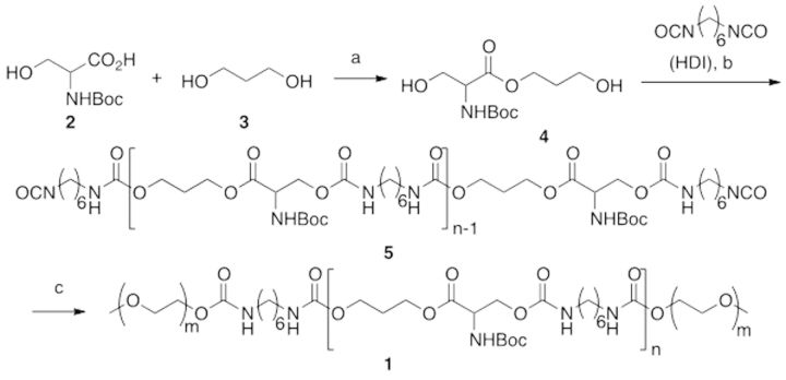
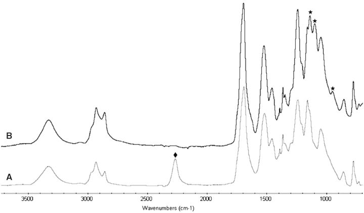
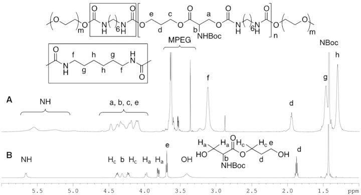
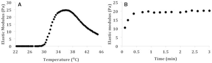
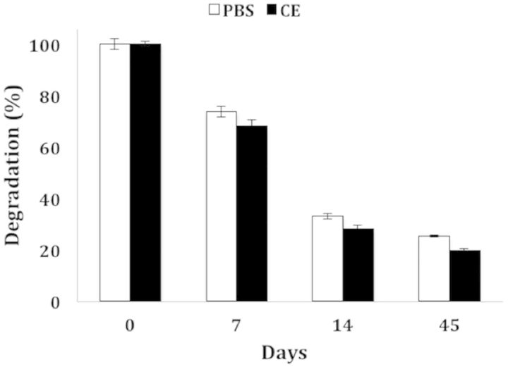
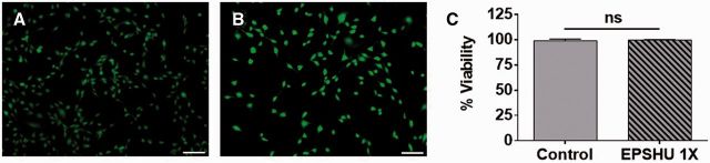
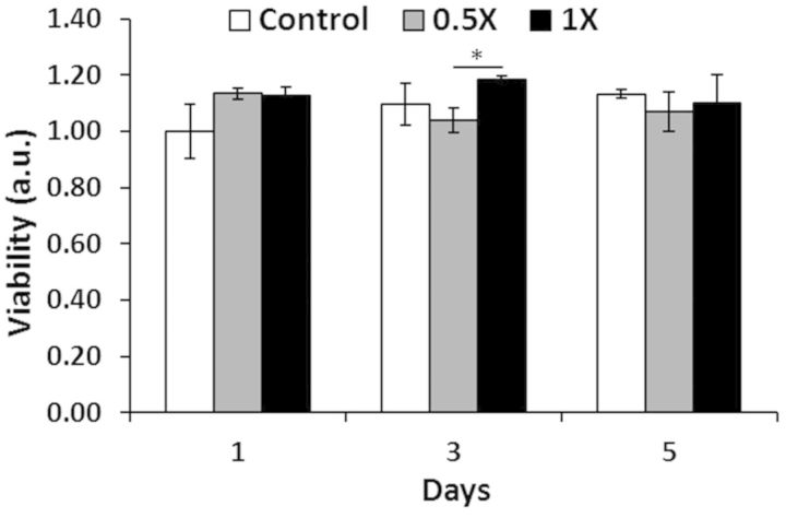
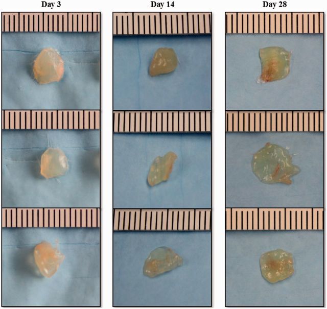
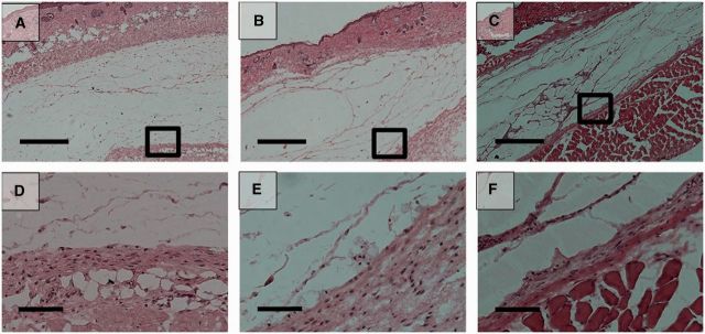
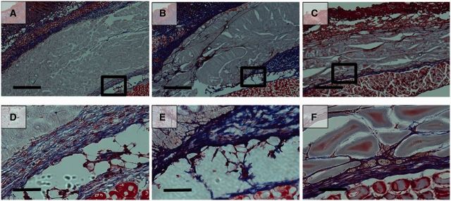
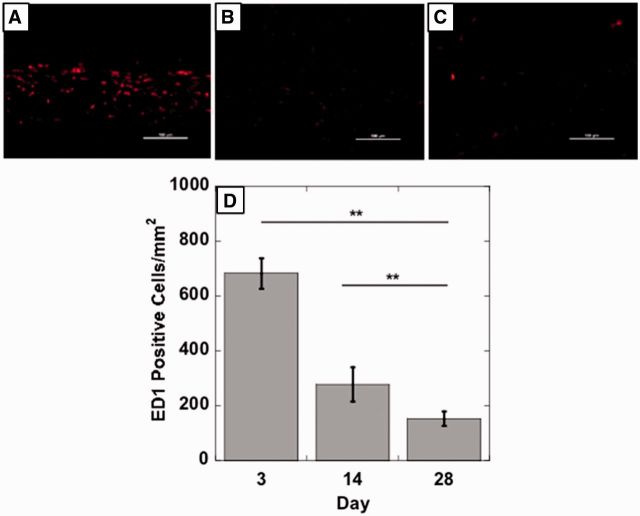
Injectable thermal gels are a useful tool for drug delivery and tissue engineering. However, most thermal gels do not solidify rapidly at body temperature (37°C). We addressed this by synthesizing a thermo-sensitive, rapidly biodegrading hydrogel. Our hydrogel, poly(ethylene glycol)-co-poly(propanol serinate hexamethylene urethane) (EPSHU), is an ABA block copolymer comprising A, methoxy poly ethylene glycol group and B, poly (propanol L-serinate hexamethylene urethane). EPSHU was characterized by gel permeation chromatography for molecular weight and (1)H NMR and Fourier transformed infrared for structure. Rheological studies measured the phase transition temperature. In vitro degradation in cholesterol esterase and in Dulbecco's phosphate buffered saline (DPBS) was tracked using the average molecular weight measured by gel permeation chromatography. LIVE/DEAD and resazurin reduction assays performed on NIH 3T3 fibroblasts exposed to EPSHU extracts demonstrated no cytotoxicity. Subcutaneous implantation into BALB/cJ mice indicated good biocompatibility in vivo. The biodegradability and biocompatibility of EPSHU together make it a promising candidate for drug delivery applications that demand carrier gel degradation within months.

Keywords: biodegradable; drug delivery; materials synthesize; thermoresponsive hydrogel.


Figures

Scheme 1.
Scheme 1.
Synthesis of EPSHU: (a) DCC, DMAP, 0 − rt, 24 h, 88%; (b) 90 °C, 2 h, then HDI in excess, DMF, 90 °C, 16 h; (c) MPEG 550, DMF, 90 °C, 24 h. 151x72mm (100 x 100 DPI).
Figure 1.
Figure 1.
FTIR spectra of (A) intermediate 5 and (B) EPSHU (1): after the PEGylation, the reactive isocyanate groups of intermediate 5 at 2267 cm−1 (diamonds) completely disappeared and peaks at 975, 1099 and 1139 cm−1 (stars) appeared.
Figure 2.
Figure 2.
1H NMR: comparison of EPSHU (1) (A) and serinate (4) (B).
Figure 3.
Figure 3.
The elastic modulus of EPSHU 10% (wt/v). (A) Temperature sweep was recorded over the range 22–45°C. (B) Time sweep was recorded at 37°C for 3 min.
Figure 4.
Figure 4.
Degradation of EPSHU in DPBS and in CE solution, expressed as percent of average MW at Day 0. The degradation rate in CE solution was barely faster than in DPBS alone. Data are presented as mean ± SD (n = 3).
Figure 5.
Figure 5.
NIH 3T3 LIVE/DEAD assay. (A) Culture media control. (B) 1X EPSHU extract. Green indicates a live cell, and red indicates a dead cell. Color images are available online. All cells are green (light gray in the black and white images), no red cells were detected. Bar is 100 µm. (C) Percent viability in 3T3s exposed to control media or 1X EPSHU extract. Data are presented as mean ± SD (n = 3).
Figure 6.
Figure 6.
CellTiter-Blue assay: effects of 0.5X and 1X strength EPSHU extracts on NIH 3T3 metabolic activity after 1, 3 and 5 days of exposure. Data are presented as mean ± SD (n = 3), normalized to the control (culture media) absorbance at Day 1. *P < 0.05.
Figure 7.
Figure 7.
EPSHU gels explanted from mice after 3, 14 and 28 days (n = 3).
Figure 8.
Figure 8.
Hematoxylin and eosin (H&E) -stained histological sections of the tissue adjacent to the sites of subcutaneous injection of 100 μl of 20% (wt/v) EPSHU into BALB/cJ mice. The EPSHU degraded gradually. Cavities with cell infiltrates had formed inside the gel at Day 3 (A and D), Day 14 (B and E) and Day 28 (C and F) after injection. (A–C) Low magnification of images (40×, scale bar = 500 μm). The rectangular frames indicate the fields chosen for capture at higher magnifications (D–F, 200×, scale bar = 100 μm).
Figure 9.
Figure 9.
MTS histological sections of the tissue adjacent to the sites of subcutaneous injection of 100 μl of 20% (wt/v) EPSHU into BALB/cJ mice. MTS confirmed that collagen deposition around the polymer (blue fibers, color images are available in downloadable pdf online.) was minimal at Day 3 (A and D), Day 14 (B and E) and Day 28 (C and F). (A–C) Low magnification of images (40×, scale bar = 500 μm). The rectangular frames indicate the fields chosen for capture at higher magnifications (D–F, 200×, scale bar = 100 μm).
Figure 10.
Figure 10.
Representative photomicrographs (200×, scale bar = 100 μm) of injection sites immunohistochemically stained with ED1, an antibody against the monocyte/macrophage marker CD68. Tissues were harvested on (A) Day 3, (B) Day 14 and (C) Day 28 post-injection. (D) The number of ED1-positive cells decreased with time, indicating a reduction in the inflammatory response. Images from 5 random areas around the injection sites were used for quantification at each time point. Data are shown as mean ± SD (n > 5). **P < 0.001.

References

    1. Mahajan HS, Tyagi V, Lohiya G, et al. Thermally reversible xyloglucan gels as vehicles for nasal drug delivery. Drug Deliv 2012;19:270–6. - PubMed
    1. Park D, Shah V, Rauck BM, et al. An anti-angiogenic reverse thermal gel as a drug-delivery system for age-related wet macular degeneration. Macromol Biosci 2013;13:464–9. - PMC - PubMed
    1. Bhosale RR, Osmani AR, Ghodake PP, et al. International Journal of Pharmaceutical and Medicinal Research. Int J Pharm Med Res 2013;1:60–9.
    1. Justin G, Guiseppi-Elie A. Characterization of electroconductive blends of poly(HEMA-co-PEGMA-co-HMMA-co-SPMA) and poly(Py-co-PyBA). Biomacromolecules 2009;10:2539–49. - PubMed
    1. Lin CC, Metters AT, Anseth KS. Functional PEG-peptide hydrogels to modulate local inflammation induced by the pro-inflammatory cytokine TNFalpha. Biomaterials 2009;30:4907–14. - PMC - PubMed

LinkOut - more resources