. 2016 Mar 18;291(12):6534-45.
doi: 10.1074/jbc.M115.696831. Epub 2016 Jan 26.

The Tumor-suppressive Small GTPase DiRas1 Binds the Noncanonical Guanine Nucleotide Exchange Factor SmgGDS and Antagonizes SmgGDS Interactions with Oncogenic Small GTPases

Affiliations

The Tumor-suppressive Small GTPase DiRas1 Binds the Noncanonical Guanine Nucleotide Exchange Factor SmgGDS and Antagonizes SmgGDS Interactions with Oncogenic Small GTPases

Carmen Bergom et al. J Biol Chem. .

Erratum in

Abstract

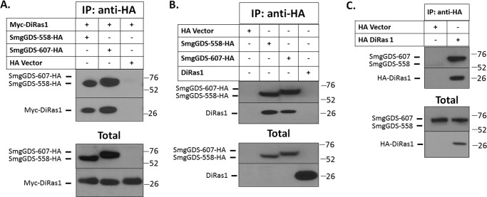
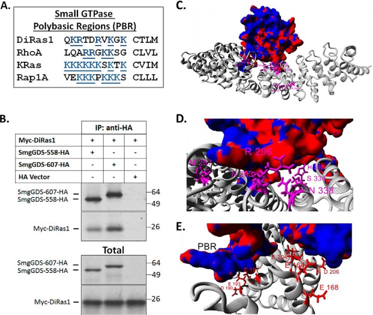
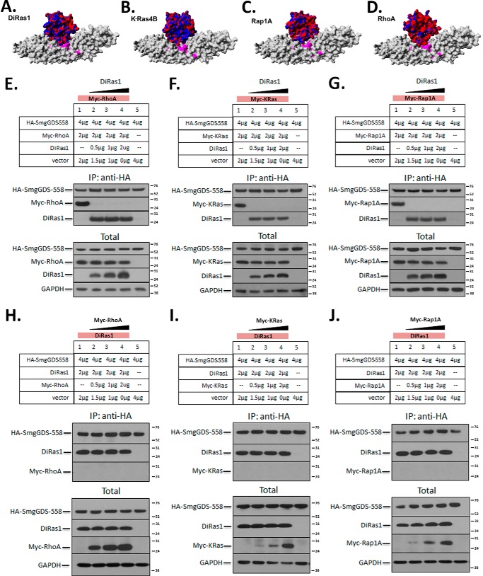
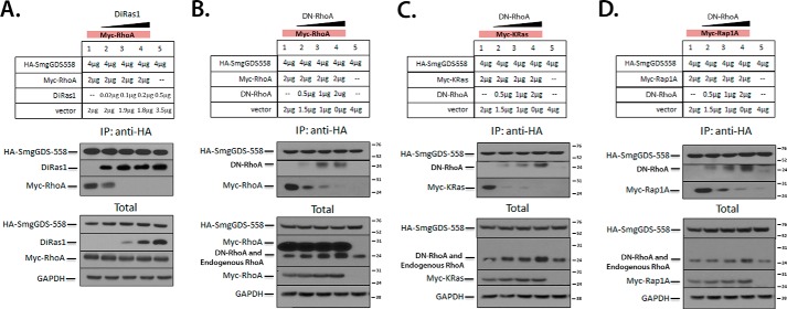
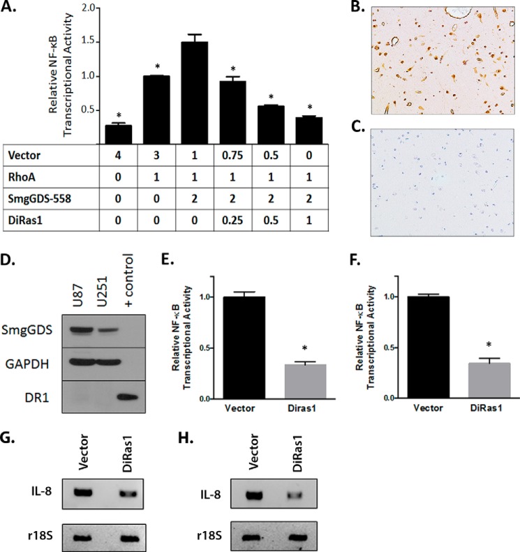
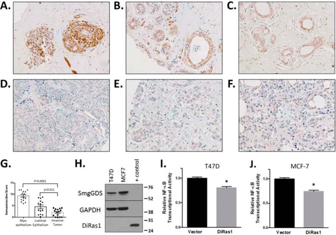
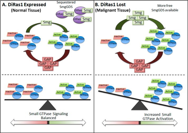
The small GTPase DiRas1 has tumor-suppressive activities, unlike the oncogenic properties more common to small GTPases such as K-Ras and RhoA. Although DiRas1 has been found to be a tumor suppressor in gliomas and esophageal squamous cell carcinomas, the mechanisms by which it inhibits malignant phenotypes have not been fully determined. In this study, we demonstrate that DiRas1 binds to SmgGDS, a protein that promotes the activation of several oncogenic GTPases. In silico docking studies predict that DiRas1 binds to SmgGDS in a manner similar to other small GTPases. SmgGDS is a guanine nucleotide exchange factor for RhoA, but we report here that SmgGDS does not mediate GDP/GTP exchange on DiRas1. Intriguingly, DiRas1 acts similarly to a dominant-negative small GTPase, binding to SmgGDS and inhibiting SmgGDS binding to other small GTPases, including K-Ras4B, RhoA, and Rap1A. DiRas1 is expressed in normal breast tissue, but its expression is decreased in most breast cancers, similar to its family member DiRas3 (ARHI). DiRas1 inhibits RhoA- and SmgGDS-mediated NF-κB transcriptional activity in HEK293T cells. We also report that DiRas1 suppresses basal NF-κB activation in breast cancer and glioblastoma cell lines. Taken together, our data support a model in which DiRas1 expression inhibits malignant features of cancers in part by nonproductively binding to SmgGDS and inhibiting the binding of other small GTPases to SmgGDS.

Keywords: Di-Ras1; GTPase Kras (KRAS); NF-kappaB (NF-KB); Rap1GDS1; Ras homolog gene family, member A (RhoA); Ras-related protein 1 (Rap1); Rig; SmgGDS; breast cancer; glioblastoma.


Figures

FIGURE 1.
FIGURE 1.
DiRas1 binds the small GTPase-modulating protein SmgGDS. A, HEK293T cells were co-transfected with SmgGDS-HA and vector or Myc-DiRas1 cDNAs and after 24 h were lysed, immunoprecipitated with anti-HA antibodies, and subjected to Western blotting with anti-HA and anti-Myc antibodies. B, HEK293T cells were transfected with cDNAs for HA-tagged SmgGDS-558 and SmgGDS-607. After 24 h, the association of endogenous DiRas1 with SmgGDS-HA was examined as in A. Western blotting of the total cell lysates (total) shows endogenous DiRas1 and HA-tagged SmgGDS. C, HA-DiRas1 was transfected into HEK293T cells, and the association of endogenous SmgGDS was examined after 24 h via immunoprecipitation as in A. Western blotting was performed for endogenous SmgGDS and HA-tagged DiRas1. The results are representative of three independent experiments.
FIGURE 2.
FIGURE 2.
DiRas1 is predicted to bind to SmgGDS in the same way that RhoA binds SmgGDS. A, the PBRs of DiRas1 and other small GTPases known to bind to SmgGDS are shown. DiRas1 has a PBR containing basic residues, similar to the other small GTPases shown. B, Myc-DiRas1 and HA-SmgGDS cDNAs were translated in the presence of [35S]methionine in in vitro transcription and translation assays. In vitro translated proteins were co-incubated and immunoprecipitated (IP) with anti-HA antibodies, and Western blotting was performed with anti-HA and anti-Myc antibodies. C–E, in silico docking studies of DiRas1 (electrostatic surface) on SmgGDS-558 (gray) suggest that DiRas1 binds to SmgGDS similarly to the way in which RhoA binds SmgGDS. The top docking conformation for DiRas1 is shown as an electrostatic surface plot (red, electronegative; blue, electropositive). SmgGDS variants known to alter RhoA guanine nucleotide exchange (4) are colored magenta (D), and electrostatic amino acids needed for RhoA binding are colored red (E). The close proximity of these amino acids to the docking interface suggests the DiRAs1 and RhoA bind to SmgGDS in a similar manner.
FIGURE 3.
FIGURE 3.
SmgGDS is not a GEF for DiRas1, and SmgGDS exhibits a higher affinity for DiRas1 than for RhoA. A and B, MANT-GTP guanine nucleotide exchange assays demonstrate that SmgGDS can mediate guanine nucleotide exchange of RhoA (A) but does not mediate guanine nucleotide exchange of DiRas1 (B). Fluorescence with exchange buffer alone or with SmgGDS (final concentration, 20 μm) or EDTA (final concentration, 10 μm) was measured. At the indicated time (arrow), RhoA or DiRas1 (2 μm) was added, and nucleotide exchange was measured. Relative fluorescent values were obtained by normalizing values to the baseline fluorescence value. C, MANT-GTP assay demonstrates that DiRas1 can inhibit guanine nucleotide exchange of RhoA in the presence of SmgGDS. Using the same methods as in A and B, fluorescence with SmgGDS (final concentration, 20 μm) was measured. For the black curve, RhoA (2 μm, black arrow) was added to SmgGDS at the indicated time. For the red curve, DiRas1 (2 μm, red arrow) was added to SmgGDS at the indicated time, and RhoA was added after an additional 60 s, as indicated (black arrow). D and E, the binding affinity of SmgGDS and RhoA (D) and SmgGDS and DiRas1 (E) was determined using a biolayer interferometry assay as described under “Experimental Procedures.” The Kd for SmgGDS and RhoA was 1.9 ± 0.1 μm, whereas the Kd for SmgGDS and DiRas1 was 39 ± 3 nm. The results are representative of two or more independent experiments.
FIGURE 4.
FIGURE 4.
DiRas1 expression potently inhibits SmgGDS association with other small GTPases. A–D, in silico docking studies suggest that DiRas1 (A, electrostatic surface) binds to SmgGDS-558 (gray surface plot) in a similar manner to K-Ras4B (B), Rap1A (C), and RhoA (D). The color scheme is as defined in Fig. 2. E–G, SmgGDS-558-HA and Myc-tagged RhoA (E), KRas-4B (F), and Rap1A (G) cDNAs were co-transfected into HEK293T cells along with increasing amounts of DiRas1 cDNA as indicated, such that all cells were transfected with identical amounts of total cDNA. After 24 h the cells were lysed, immunoprecipitated with anti-HA antibodies, and subjected to Western blotting for HA and Myc tags, as well as DiRas1 and GAPDH (lysates). DiRas1 expression inhibited SmgGDS association with all other small GTPases examined. The results are representative of three independent experiments. H–J, SmgGDS-558-HA and increasing amounts of Myc-tagged RhoA (H), KRas-4B (I), and Rap1A (J) cDNAs were co-transfected into HEK293T cells along with a constant level of DiRas1 cDNA as indicated, such that all cells were transfected with identical amounts of total cDNA. After 24 h, the cells were lysed, immunoprecipitated with anti-HA antibodies, and subjected to Western blotting as described in E. The results are representative of two or more independent experiments.
FIGURE 5.
FIGURE 5.
Diras1 potently inhibits binding of RhoA to SmgGDS and acts in a fashion similar to DN-RhoA. A, co-transfection of tagged SmgGDS and RhoA in HEK293T cells was performed as in Fig. 4E, with much lower levels of increasing untagged DiRas1 (amounts indicated), such that all cells were transfected with identical amounts of total cDNA. After 24 h the cells were lysed, immunoprecipitated with anti-HA antibodies, and subjected to Western blotting for HA and Myc tags, as well as DiRas1 and GAPDH (lysates). B–D, DN-RhoA inhibits small GTPase binding to SmgGDS in a manner similar to WT DiRas1. Co-transfections of HEK293T cells were performed as indicated in Fig. 4E, with constant levels of Myc-tagged RhoA (B), KRas (C), and Rap1A (D), and increasing levels of untagged DN-RhoA, as indicated. The cells were lysed after 24 h, and immunoprecipitation with anti-HA antibodies and Western blotting were performed as follows (top to bottom in each section): anti-HA, anti-RhoA (showing immunoprecipitated untagged DN-RhoA), and anti-Myc. In the lysate panels, from top to bottom, antibodies used were: anti-HA, untagged RhoA (which binds tagged RhoA in B, untagged DN-RhoA, and endogenous RhoA, as indicated), Myc tag, and GAPDH. The results are representative of two or more independent experiments.
FIGURE 6.
FIGURE 6.
DiRas1 inhibits RhoA- and SmgGDS-mediated NF-κB transcriptional activity. A, DiRas1 expression mitigates RhoA- and SmgGDS-induced NF-κB activation. HEK293T cells were co-transfected with NF-κB reporter plasmid, along with cDNAs encoding RhoA, SmgGDS-558, DiRas1, and empty vector in the amounts indicated in μg. After 24 h, luminescence was quantified and normalized, with RhoA-mediated activation set as 1.0. The results are from three independent experiments. *, p < 0.0001 via one-way analysis of variance with Dunnett's multiple comparison tests, when compared with the cells expressing SmgGDS and RhoA. B and C, IHC staining of nonmalignant human cortex tissue reveals DiRas1 staining (B) in glial cells and other cell types (20× magnification), with no staining using an isotype control (C). D, glioblastoma U87 and U251 cells lines (50 μg of lysate each) lack detectable DiRas1 protein expression. Control cell lysate (3 μg) is HEK293T cells expressing DiRas1 cDNA. E and F, re-expressing DiRas1 in glioblastoma cell lines reduces basal NF-κB transcriptional activity. U87 (E) and U251 (F) cells were transfected with empty vector or DiRas1 and NF-κB reporter and β-gal. After 24 h, luminescence was quantified and normalized. The results are the means ± S.E. from at least three independent experiments. *, p < 0.0001 compared with vector-transfected cells using the Mann-Whitney U test. G and H, mRNA expression of the NF-κB regulated gene IL-8 is decreased in U87 (G) and U251 (H) cells expressing DiRas1 cDNA, compared with vector-transfected cells. The cells were transfected with vector or DiRas1 cDNA for 72 h, total RNA was extracted and reverse transcribed to cDNA, and RT-PCR was performed, using r18S as a control. The results are representative of three independent experiments.
FIGURE 7.
FIGURE 7.
DiRas1 is expressed in normal breast epithelium but absent from breast cancers, and it inhibits NF-κB transcriptional activity in breast cancer cell lines. A–F, DiRas1 is expressed in normal human ductal epithelium and myoepithelium (A–C) but is expressed to a lesser extent in most invasive breast tumors (D–F). These images are representative of the samples quantified in Table 1. G, scatter dot plot shows the IRS score for each sample, as well as the means ± the S.E. H, the human breast cancer cell lines T47D and MCF-7 (50 μg each) express no detectable DiRas1 protein, respectively. Control cell lysate (4 μg) is HEK293T cells expressing DiRas1 cDNA. I and J, DiRas1 expression diminishes basal NF-κB transcriptional activity in T47D (I) and MCF-7 (J) cells. The breast cancer cells were transfected with empty vector or DiRas1 and NF-κB reporter and β-gal. After 24 h, luminescence was quantified and normalized to vector-transfected cells. The results are the means ± S.E. from at least three independent experiments. *, p < 0.0001 compared with vector-transfected cell activity using the Mann-Whitney U test.
FIGURE 8.
FIGURE 8.
Proposed model of DiRas1-mediated alteration in oncogenic small GTP signaling via binding to SmgGDS. A, in noncancerous tissues where DiRas1 is expressed, it can nonproductively bind SmgGDS and diminish SmgGDS interactions with other small GTPases, promoting balanced oncogenic small GTPase activity. B, in malignant tissues when DiRas1 expression is decreased or lost, more SmgGDS protein is available to interact with oncogenic small GTPases and promote their activation, resulting in increased pro-oncogenic small GTPase signaling.

References

    1. Alan J. K., and Lundquist E. A. (2013) Mutationally activated Rho GTPases in cancer. Small GTPases 4, 159–163 - PMC - PubMed
    1. Bos J. L. (1989) ras oncogenes in human cancer: a review. Cancer Res. 49, 4682–4689 - PubMed
    1. Pylayeva-Gupta Y., Grabocka E., and Bar-Sagi D. (2011) RAS oncogenes: weaving a tumorigenic web. Nat. Rev. Cancer 11, 761–774 - PMC - PubMed
    1. Hamel B., Monaghan-Benson E., Rojas R. J., Temple B. R., Marston D. J., Burridge K., and Sondek J. (2011) SmgGDS is a guanine nucleotide exchange factor that specifically activates RhoA and RhoC. J. Biol. Chem. 286, 12141–12148 - PMC - PubMed
    1. Berg T. J., Gastonguay A. J., Lorimer E. L., Kuhnmuench J. R., Li R., Fields A. P., and Williams C. L. (2010) Splice variants of SmgGDS control small GTPase prenylation and membrane localization. J. Biol. Chem. 285, 35255–35266 - PMC - PubMed

Publication types

MeSH terms

Associated data

LinkOut - more resources