Comparative Study
. 2015 Nov-Dec;23(6):599-608.
doi: 10.1590/1678-775720150122.

Cytocompatibility and biocompatibility of nanostructured carbonated hydroxyapatite spheres for bone repair

Affiliations
Comparative Study

Cytocompatibility and biocompatibility of nanostructured carbonated hydroxyapatite spheres for bone repair

Mônica Diuana Calasans-Maia et al. J Appl Oral Sci. 2015 Nov-Dec.

Abstract

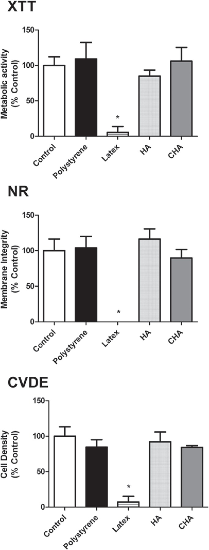
Objective: The aim of this study was to investigate the in vitro and in vivo biological responses to nanostructured carbonated hydroxyapatite/calcium alginate (CHA) microspheres used for alveolar bone repair, compared to sintered hydroxyapatite (HA).

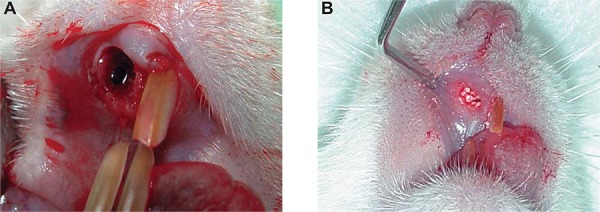
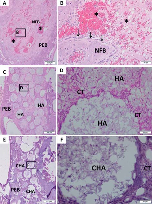
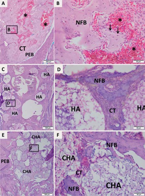
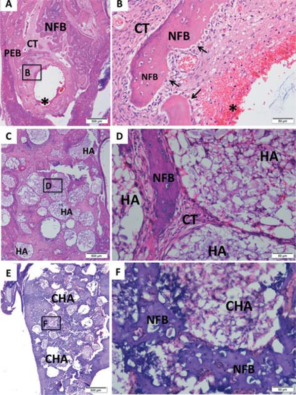
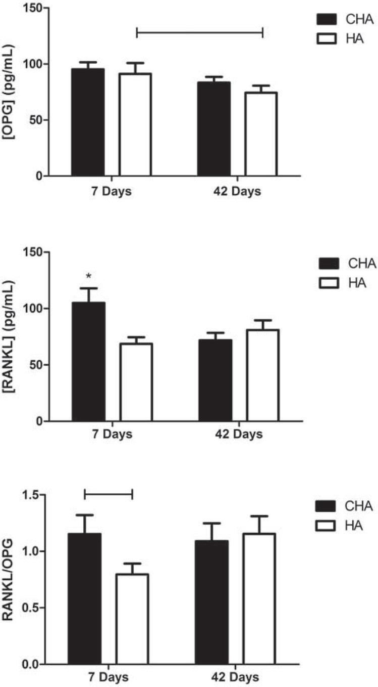
Material and methods: The maxillary central incisors of 45 Wistar rats were extracted, and the dental sockets were filled with HA, CHA, and blood clot (control group) (n=5/period/group). After 7, 21 and 42 days, the samples of bone with the biomaterials were obtained for histological and histomorphometric analysis, and the plasma levels of RANKL and OPG were determined via immunoassay. Statistical analysis was performed by Two-Way ANOVA with post-hoc Tukey test at 95% level of significance.

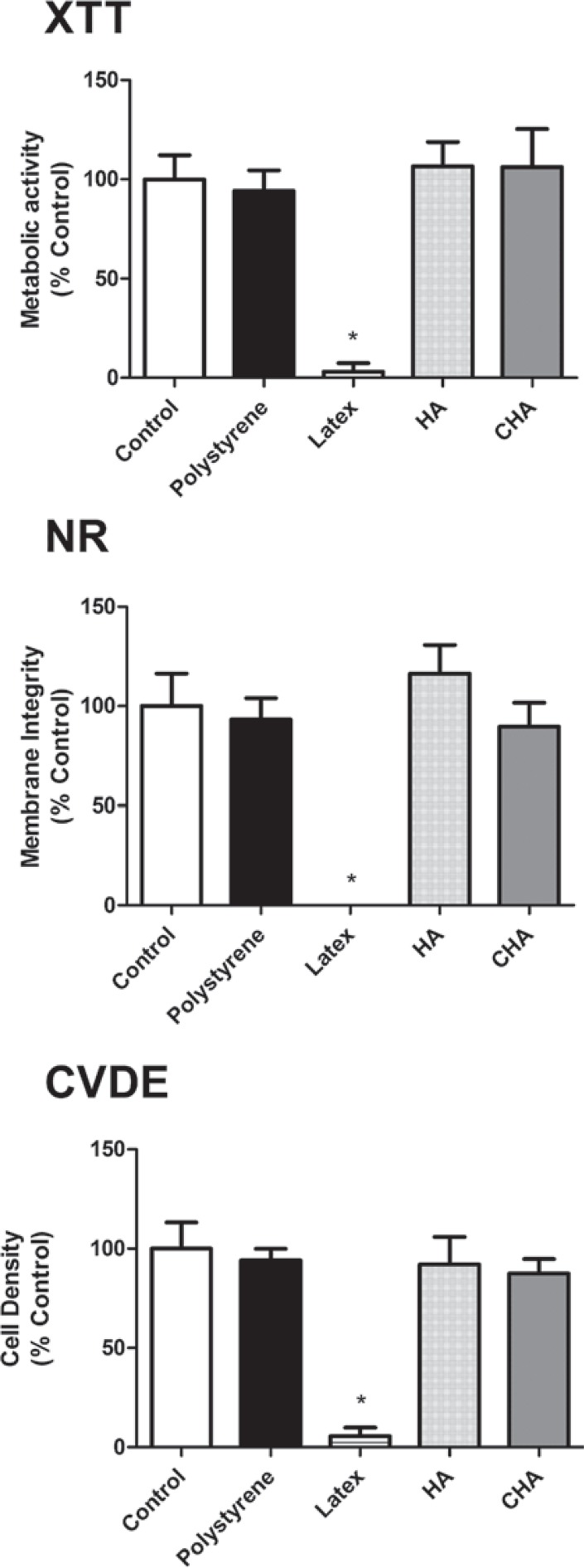
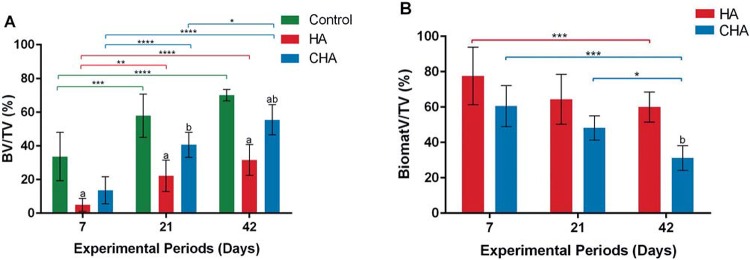
Results: The CHA and HA microspheres were cytocompatible with both human and murine cells on an in vitro assay. Histological analysis showed the time-dependent increase of newly formed bone in control group characterized by an intense osteoblast activity. In HA and CHA groups, the presence of a slight granulation reaction around the spheres was observed after seven days, which was reduced by the 42nd day. A considerable amount of newly formed bone was observed surrounding the CHA spheres and the biomaterials particles at 42-day time point compared with HA. Histomorphometric analysis showed a significant increase of newly formed bone in CHA group compared with HA after 21 and 42 days from surgery, moreover, CHA showed almost 2-fold greater biosorption than HA at 42 days (two-way ANOVA, p<0.05) indicating greater biosorption. An increase in the RANKL/OPG ratio was observed in the CHA group on the 7th day.

Conclusion: CHA spheres were osteoconductive and presented earlier biosorption, inducing early increases in the levels of proteins involved in resorption.


Figures

Figure 1
Figure 1. X-Ray Diffraction pattern (XRD) of the carbonated hydroxyapatite (CHA) powder. Observe broad peaks due to the nanostructured and poorly crystalline character of the CHA
Figure 2
Figure 2. Scanning Electron Microscopy (SEM) micrographs. A: Carbonated hydroxyapatite (CHA) spheres; B: CHA spheres surface, both without thermal treatment; C: Hydroxyapatite (HA) spheres, and D: HA spheres surface. A and C: 100X magnification, and B and D: 1,000X magnification. Scale bars of 1mm (A and C) and 100 µm (B and D)
Figure 3
Figure 3. Surgical procedures for biomaterials implantation: A: The maxillary right incisor was extracted, and B: The socket was filled with spheres of biomaterials according to the experimental group.
Figure 4
Figure 4. Multiparametric cytotoxicity assay with murine MC3T3-E1 cells. Results showed as percentage of control group (Control; exposure to DMEM). * ≠ all the other groups (p<0.05)
Figure 5
Figure 5. Multiparametric cytotoxicity assay with human osteoblastic cells. Results showed as percentage of control group (Control; exposure to DMEM). * ≠ all the other groups (p<0.05)
Figure 6
Figure 6. Representative photomicrographs of alveolar socket after 7 days. A and B: Control group; C and D: HA group; E and F: CHA group. The small squares identified by B, D and F are displayed at 40-fold magnification adjacent to the figures with lower magnification (Figures A, C and E, respectively). Pre-existing bone (PEB); Connective tissue (CT); Osteoblasts pavement (black arrow); serum hemorrhagic exudate (*); Newly formed bone (NFB); Neutrophilic infiltrate (n); Osteoid (o); hydroxyapatite (HA); nanostructured carbonated hydroxyapatite/calcium alginate (CHA). Hematoxylin and eosin stained.
Figure 7
Figure 7. Representative photomicrographs of alveolar socket after 21 days. A and B: Control group; C and D: HA group; E and F: CHA group. The small squares identified by B, D and F are displayed at 40-fold magnification adjacent to the figures with lower magnification (Figures A, C and E, respectively). Pre-existing bone (PEB); Connective tissue (CT); Osteoblasts pavement (black arrow); hemorrhagic exudate (*); Newly formed bone (NFB); hydroxyapatite (HA); nanostructured carbonated hydroxyapatite/calcium alginate (CHA). Hematoxylin and eosin stained
Figure 8
Figure 8. Representative photomicrographs of alveolar socket after 42 days. A and B: Control group; C and D: HA group; E and F: CHA group. The small squares identified by B, D and F are displayed at 40-fold magnification adjacent to the figures with lower magnification (Figures A, C and E, respectively). Pre-existing bone (PEB); Connective tissue (CT); Osteoblasts pavement (black arrow); hemorrhagic exudate (*); Newly formed bone (NFB); hydroxyapatite (HA); nanostructured carbonated hydroxyapatite/calcium alginate (CHA). Hematoxylin and eosin stained
Figure 9
Figure 9. The bone volume density (BV/TV%) (A), and biomaterials volume density (BiomatV/TV%) (B) after 7, 21, and 42 days of implantation. (a) significant statistical differences between hydroxyapatite (HA) or nanostructured carbonated hydroxyapatite/calcium alginate (CHA) compared with control group at the same experimental period; (b) significant statistical differences between CHA and HA at the same experimental period; (*) significant statistical difference between the same group at different experimental periods (two-way ANOVA, p<0.05). Results are shown as mean percentages±confidence interval
Figure 10
Figure 10. Serum concentrations assay of RANKL/OPG in operated rats after 7 and 42 days. Abbreviations: CHA=carbonated hydroxyapatite/sodium alginate; HA=stoichiometry hydroxyapatite. Bar ≠ between groups (p<0.05); * ≠ all the other groups (p<0.05)

References

    1. Almasri M, Altalibi M. Efficacy of reconstruction of alveolar bone using an alloplastic hydroxyapatite tricalcium phosphate graft under biodegradable chambers. Br J Oral Maxillofac Surg. 2011;49:469–473. - PubMed
    1. Alves Cardoso D, Jansen JA, Leeuwenburgh SC. Synthesis and application of nanostructured calcium phosphate ceramics for bone regeneration. J Biomed Mater Res B Appl Biomater. 2012;100:2316–2326. - PubMed
    1. Bauer TW. Bone graft substitutes. Skeletal Radiol. 2007;36:1105–1107. - PubMed
    1. Brandt J, Henning S, Michler G, Hein W, Bernstein A, Schulz M. Nanocrystalline hydroxyapatite for bone repair: an animal study. J Mater Sci Mater Med. 2010;21:283–294. - PubMed
    1. Calasans-Maia M, Calasans-Maia J, Santos S, Mavropoulos E, Farina M, Lima I, et al. Short-term in vivo evaluation of zinc-containing calcium phosphate using a normalized procedure. Mater Sci Eng C Mater Biol Appl. 2014;41:309–319. - PubMed

Publication types

MeSH terms