. 2016 Jul;55(1):135-49.
doi: 10.1165/rcmb.2015-0327OC.

The 78-kD Glucose-Regulated Protein Regulates Endoplasmic Reticulum Homeostasis and Distal Epithelial Cell Survival during Lung Development

Affiliations

The 78-kD Glucose-Regulated Protein Regulates Endoplasmic Reticulum Homeostasis and Distal Epithelial Cell Survival during Lung Development

Per Flodby et al. Am J Respir Cell Mol Biol. 2016 Jul.

Abstract

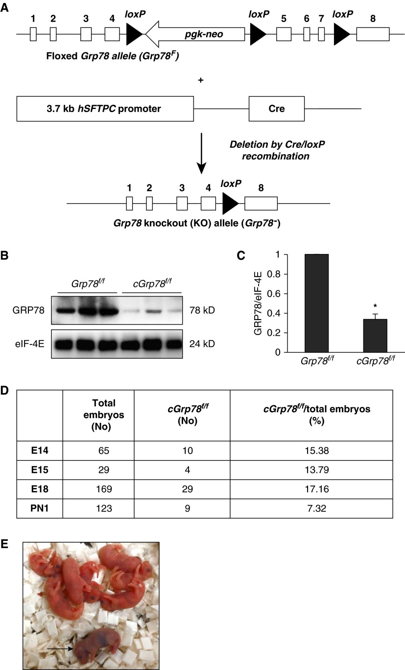
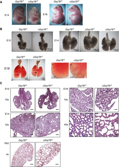
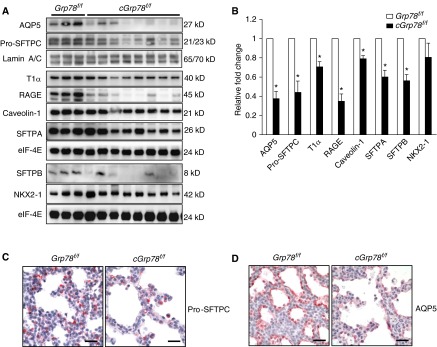
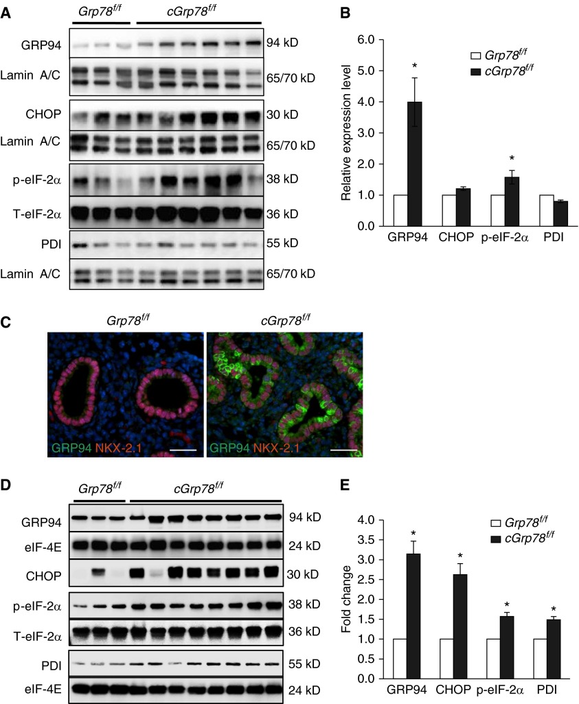
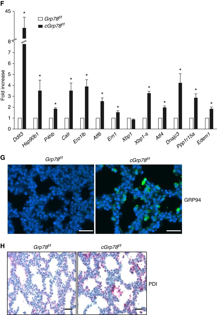
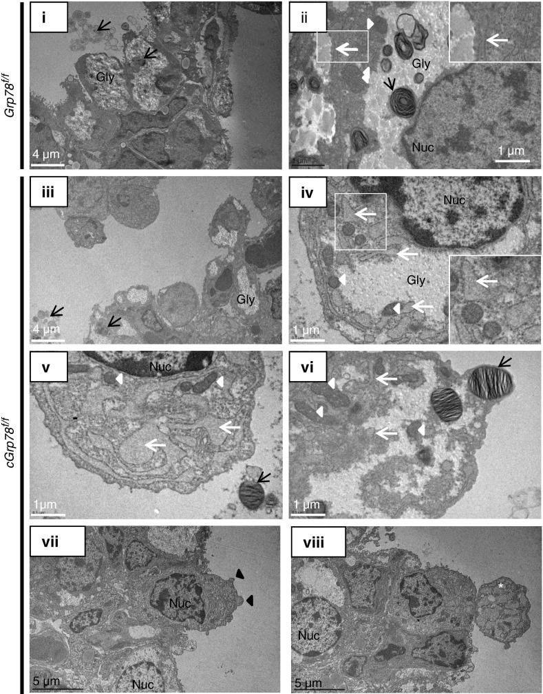
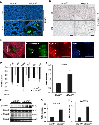
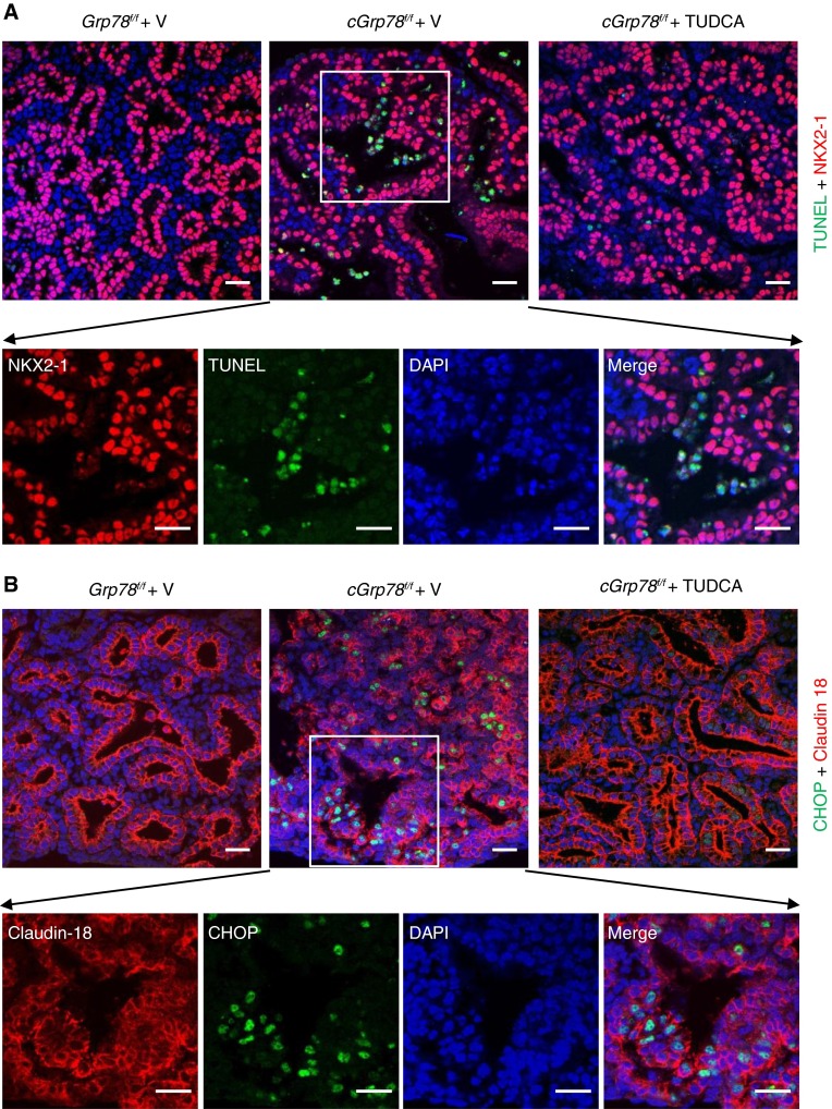
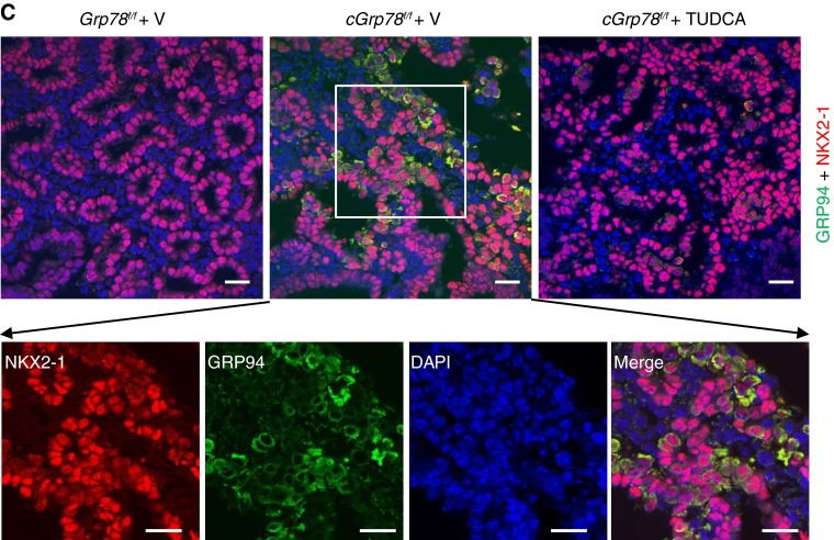
Bronchopulmonary dysplasia (BPD), a chronic lung disease of prematurity, has been linked to endoplasmic reticulum (ER) stress. To investigate a causal role for ER stress in BPD pathogenesis, we generated conditional knockout (KO) mice (cGrp78(f/f)) with lung epithelial cell-specific KO of Grp78, a gene encoding the ER chaperone 78-kD glucose-regulated protein (GRP78), a master regulator of ER homeostasis and the unfolded protein response (UPR). Lung epithelial-specific Grp78 KO disrupted lung morphogenesis, causing developmental arrest, increased alveolar epithelial type II cell apoptosis, and decreased surfactant protein and type I cell marker expression in perinatal lungs. cGrp78(f/f) pups died immediately after birth, likely owing to respiratory distress. Importantly, Grp78 KO triggered UPR activation with marked induction of the proapoptotic transcription factor CCAAT/enhancer-binding proteins (C/EBP) homologous protein (CHOP). Increased expression of genes involved in oxidative stress and cell death and decreased expression of genes encoding antioxidant enzymes suggest a role for oxidative stress in alveolar epithelial cell (AEC) apoptosis. Increased Smad3 phosphorylation and expression of transforming growth factor-β/Smad3 targets Cdkn1a (encoding p21) and Gadd45a suggest that interactions among the apoptotic arm of the UPR, oxidative stress, and transforming growth factor-β/Smad signaling pathways contribute to Grp78 KO-induced AEC apoptosis and developmental arrest. Chemical chaperone Tauroursodeoxycholic acid reduced UPR activation and apoptosis in cGrp78(f/f) lungs cultured ex vivo, confirming a role for ER stress in observed AEC abnormalities. These results demonstrate a key role for GRP78 in AEC survival and gene expression during lung development through modulation of ER stress, and suggest the UPR as a potential therapeutic target in BPD.

Keywords: 78-kD glucose-regulated protein knockout; alveolar epithelial cells; apoptosis; endoplasmic reticulum stress/unfolded protein response signaling.


Figures

Figure 1.
Figure 1.
Embryonic and early perinatal lethality in chaperone 78-kD glucose-regulated protein (cGrp78f/f) mice. (A) Schematic diagram of floxed Grp78 (Grp78f) and knockout (KO) alleles (Grp78). Western blotting (WB) (B) and corresponding quantitative analysis (C) show significantly decreased GRP78 expression in cGrp78f/f whole-lung lysates at Embryonic Day 18 (E18). Eukaryotic translation Initiation factor 4E (eIF-4E) was used as loading control. *P < 0.05, n = 3. (D) The number of cGrp78f/f embryos and live births at Postnatal Day 1 (PN1) was less than the expected Mendelian ratio of 25%. (E) cGrp78f/f mice (arrow) died within a few hours after birth. hSFTPC, human surfactant protein C.
Figure 2.
Figure 2.
Abnormal lung morphology in cGrp78f/f mice. (A) Embryos appear macroscopically normal at both E14 and E18. (B) Lungs are morphologically normal in cGrp78f/f embryos at E12 but display dilated distal airspaces at E14 and E18. Right panels for E18 are magnified insets of the left panels A and B, showing clear dilation. Lungs of cGrp78f/f embryos also appear smaller at E18. (C) Hematoxylin and eosin staining shows normal lung morphology at E12, with dilated distal airways by E14. At E18, airspace enlargement, irregular airspace size, and cell debris in the airspaces is evident in cGrp78f/f but not in Grp78f/f mice. At PN1, lungs display alveolar hypoplasia, alveolar airspace enlargement, and thin mesenchyme. Scale bars: 200 μm for E12, E14, and E18 at 10× magnification, 50 μm at 40× magnification, and 500 μm for PN1.
Figure 3.
Figure 3.
KO of Grp78 reduces expression of alveolar epithelial type (AT) 2 and AT1 cell markers. Representative WB using whole-lung lysates (A) and corresponding quantitative analysis (B) show decreased expression of AT2 and AT1 cell markers in cGrp78f/f mice at E18 (n ≥ 3, *P < 0.05). Immunohistochemistry confirmed decreased expression of pro-SFTPC (red) (C) and AQP5 (red) (D) in cGrp78f/f mice at E18. Scale bars: 50 μm. AQP5, Aquaporin5; NKX2-1, NK2 homeobox 1; RAGE, receptor for advanced glycation endproducts; SFTPA, surfactant protein A; SFTPB, surfactant protein B.
Figure 4.
Figure 4.
Unfolded protein response activation in lungs of cGrp78f/f mice. Representative WB using whole-lung lysates (A) and corresponding quantitative analysis (B) show significant increases of GRP94 and p-eiF2α protein in E14 lungs of cGrp78f/f mice (n ≥ 3, *P < 0.05). (C) Immunostaining demonstrates increased GRP94 expression in NKX2-1+ epithelial cells of Grp78 KO cGrp78f/f mice at E14. Scale bars: 50 μm. Representative WB (D) and corresponding quantitative analysis (E) using whole-lung lysates shows significant increases of GRP94, CCAAT/enhancer-binding proteins (C/EBP) homologous protein (CHOP), p-eiF2α, and protein disulfide isomerase (PDI) in E18 lung of cGrp78f/f mice (n ≥ 3, *P < 0.05). (F) Quantitative RT-PCR (qRT-PCR) shows increased mRNA expression of Hsp90b1, Ddit3, and P4 hb (encoding GRP94, CHOP, and PDI), and other endoplasmic reticulum (ER) stress markers in E18 lungs of cGrp78f/f mice (n = 3, *P < 0.05). Immunostaining demonstrates increased GRP94 (green) (G) and PDI (red) (H) expression in epithelial cells lining the airspaces in cGrp78f/f mice at E18. Scale bars: 50 μm.
Figure 4.
Figure 4.
Unfolded protein response activation in lungs of cGrp78f/f mice. Representative WB using whole-lung lysates (A) and corresponding quantitative analysis (B) show significant increases of GRP94 and p-eiF2α protein in E14 lungs of cGrp78f/f mice (n ≥ 3, *P < 0.05). (C) Immunostaining demonstrates increased GRP94 expression in NKX2-1+ epithelial cells of Grp78 KO cGrp78f/f mice at E14. Scale bars: 50 μm. Representative WB (D) and corresponding quantitative analysis (E) using whole-lung lysates shows significant increases of GRP94, CCAAT/enhancer-binding proteins (C/EBP) homologous protein (CHOP), p-eiF2α, and protein disulfide isomerase (PDI) in E18 lung of cGrp78f/f mice (n ≥ 3, *P < 0.05). (F) Quantitative RT-PCR (qRT-PCR) shows increased mRNA expression of Hsp90b1, Ddit3, and P4 hb (encoding GRP94, CHOP, and PDI), and other endoplasmic reticulum (ER) stress markers in E18 lungs of cGrp78f/f mice (n = 3, *P < 0.05). Immunostaining demonstrates increased GRP94 (green) (G) and PDI (red) (H) expression in epithelial cells lining the airspaces in cGrp78f/f mice at E18. Scale bars: 50 μm.
Figure 5.
Figure 5.
Grp78 KO alters ER structure. Representative electron micrographs of lung at E18 show that epithelial cells are not tightly connected, and some are detaching from the epithelium (iii) and ER (white arrows) is abnormally expanded (iv, v, and vi) in cGrp78f/f compared with control Grp78f/f mice (i and ii). Lamellar bodies (black arrows) are observed within cells and in airspaces in cGrp78f/f mice (iii and vi) and Grp78f/f mice, but they are seldom observed in cells with greater ER expansion (iv and v). More mitochondria (white arrowheads) and no increase in autophagosomes are observed in cGrp78f/f mice. Marginalization and condensation of chromatin in the nucleus (iv, vii, and viii), cell surface blebbing (black arrowheads; vii) and possible apoptotic bodies (white star; viii) are observed in cGrp78f/f mice. Gly, Glycogen; Nuc, nucleus.
Figure 6.
Figure 6.
Grp78 KO results in apoptosis, activation of oxidative stress and transforming growth factor (TGF)-β signaling in alveolar epithelial cell (AEC). (A) Terminal deoxynucleotidyl transferase dUTP nick end labeling (TUNEL) assay and (B) immunohistochemistry for cleaved (CL)-caspase 3 demonstrate increased numbers of TUNEL+ cells (green) and CL-caspase 3+ cells (brown) located in the airspaces in E18, but not E14, cGrp78f/f embryos. 4′,6-diamidino-2-phenylindole (DAPI) or methyl green is the nuclear counterstain. Scale bars: 50 μm. (C) Double immunofluorescence staining shows colocalization of CL-caspase 3 and NKX2-1 in cells detaching from the epithelial layer in cGrp78f/f lung. The three panels to the right are individual channels showing higher magnification of the rectangular area in the left image. Arrowheads shows CL-caspase 3+/NKX2-1+ cells. Scale bars: 50 μm. qRT-PCR using whole-lung RNA harvested from cGrp78f/f and Grp78f/f mice at E18 shows that Grp78 KO reduces expression of genes encoding antioxidant enzymes (D) and increases expression of Noxa1, a gene involved in generation of reactive oxidant species (E) (n = 3, *P < 0.05). (F) WB demonstrates that Grp78 KO increases p-Smad3, but not p-Smad2, expression. qRT-PCR shows increased expression of Cdkn1a (encoding p21) (G) and Gadd45a (H) in cGrp78f/f mice (n = 3–4, *P < 0.05).
Figure 7.
Figure 7.
Tauroursodeoxycholic acid (TUDCA) inhibits AEC apoptosis in cGrp78f/f lung. (A) Staining for NKX2-1 (red) and TUNEL (green) shows increased TUNEL+ cells in cGrp78f/f (middle panel) compared with Grp78f/f lung (left panel). TUNEL+ cells in cGrp78f/f lung were decreased by TUDCA treatment (right panel). Scale bar: 20 μm. The four lower panels are individual and merged channels showing higher magnification of the rectangular area in the upper middle image. Scale bars: 20 μm. (B) Staining for tight junction protein claudin 18 (red) and CHOP (green) show increased CHOP+ cells in cGrp78f/f (middle panel) compared with Grp78f/f lung (left panel). CHOP+ cells in cGrp78f/f lung were decreased by TUDCA treatment (right panel). Scale bar: 20 μm. The four lower panels are individual and merged channels showing higher magnifications of the rectangular area in the upper middle image. Scale bar: 20 μm. (C) Staining for NKX2-1 (red) and GRP94+ (green) shows increased GRP94+ cells in cGrp78f/f lung (middle panel) compared with Grp78f/f lung (left panel). GRP94+ cells in cGrp78f/f lung were decreased by TUDCA treatment (right panel). Scale bars: 20 μm. The four lower panels are individual and merged channels showing higher magnifications of the rectangular area in the upper middle image. Scale bars: 20 μm.
Figure 7.
Figure 7.
Tauroursodeoxycholic acid (TUDCA) inhibits AEC apoptosis in cGrp78f/f lung. (A) Staining for NKX2-1 (red) and TUNEL (green) shows increased TUNEL+ cells in cGrp78f/f (middle panel) compared with Grp78f/f lung (left panel). TUNEL+ cells in cGrp78f/f lung were decreased by TUDCA treatment (right panel). Scale bar: 20 μm. The four lower panels are individual and merged channels showing higher magnification of the rectangular area in the upper middle image. Scale bars: 20 μm. (B) Staining for tight junction protein claudin 18 (red) and CHOP (green) show increased CHOP+ cells in cGrp78f/f (middle panel) compared with Grp78f/f lung (left panel). CHOP+ cells in cGrp78f/f lung were decreased by TUDCA treatment (right panel). Scale bar: 20 μm. The four lower panels are individual and merged channels showing higher magnifications of the rectangular area in the upper middle image. Scale bar: 20 μm. (C) Staining for NKX2-1 (red) and GRP94+ (green) shows increased GRP94+ cells in cGrp78f/f lung (middle panel) compared with Grp78f/f lung (left panel). GRP94+ cells in cGrp78f/f lung were decreased by TUDCA treatment (right panel). Scale bars: 20 μm. The four lower panels are individual and merged channels showing higher magnifications of the rectangular area in the upper middle image. Scale bars: 20 μm.

Similar articles

Cited by

References

    1. Malhotra JD, Kaufman RJ. The endoplasmic reticulum and the unfolded protein response. Semin Cell Dev Biol. 2007;18:716–731. - PMC - PubMed
    1. Ni M, Lee AS. ER chaperones in mammalian development and human diseases. FEBS Lett. 2007;581:3641–3651. - PMC - PubMed
    1. Luo B, Lee AS. The critical roles of endoplasmic reticulum chaperones and unfolded protein response in tumorigenesis and anticancer therapies. Oncogene. 2013;32:805–818. - PMC - PubMed
    1. Ron D, Walter P. Signal integration in the endoplasmic reticulum unfolded protein response. Nat Rev Mol Cell Biol. 2007;8:519–529. - PubMed
    1. Song B, Scheuner D, Ron D, Pennathur S, Kaufman RJ. Chop deletion reduces oxidative stress, improves β cell function, and promotes cell survival in multiple mouse models of diabetes. J Clin Invest. 2008;118:3378–3389. - PMC - PubMed

MeSH terms