. 2016 Jan 27;11(1):e0147906.
doi: 10.1371/journal.pone.0147906. eCollection 2016.

Transplant Antennae and Host Brain Interact to Shape Odor Perceptual Space in Male Moths

Affiliations

Transplant Antennae and Host Brain Interact to Shape Odor Perceptual Space in Male Moths

Seong-Gyu Lee et al. PLoS One. .

Abstract

Behavioral responses to odors rely first upon their accurate detection by peripheral sensory organs followed by subsequent processing within the brain's olfactory system and higher centers. These processes allow the animal to form a unified impression of the odor environment and recognize combinations of odorants as single entities. To investigate how interactions between peripheral and central olfactory pathways shape odor perception, we transplanted antennal imaginal discs between larval males of two species of moth Heliothis virescens and Heliothis subflexa that utilize distinct pheromone blends. During metamorphic development olfactory receptor neurons originating from transplanted discs formed connections with host brain neurons within olfactory glomeruli of the adult antennal lobe. The normal antennal receptor repertoire exhibited by males of each species reflects the differences in the pheromone blends that these species employ. Behavioral assays of adult transplant males revealed high response levels to two odor blends that were dissimilar from those that attract normal males of either species. Neurophysiological analyses of peripheral receptor neurons and central olfactory neurons revealed that these behavioral responses were a result of: 1. the specificity of H. virescens donor olfactory receptor neurons for odorants unique to the donor pheromone blend and, 2. central odor recognition by the H. subflexa host brain, which typically requires peripheral receptor input across 3 distinct odor channels in order to elicit behavioral responses.


Conflict of interest statement

Competing Interests: The authors have declared that no competing interests exist.

Figures

Fig 1
Fig 1. Organization of the peripheral olfactory system in adult H. virescens and H. subflexa.
Pheromone sensitive olfactory receptor neurons (ORNs) are housed in three types of sensilla on the antennae of male H. virescens and H. subflexa. ORN axons project to antennal lobe glomeruli that constitute the macroglomerular complex (MGC) as well as part of a posterior glomerular complex (PCx). Behavioral preferences of males of the two species are accounted for by differences in the odorant affinites of olfactory receptor neurons in the B- and C-type sensilla in addition to higher order processing of these inputs. ORN affinities for odorants in brackets are weaker. An asterisk by individual ORNs indicates that the odorants associated with three pathways have behavioral significance. These pathways connect to three glomeruli in the MGC (cumulus, DM and AM, each also demarcated by an asterisk). The fourth MGC glomerulus (VM) is activated by odorants that have relatively little influence on behavior. The anatomy of the macroglomerular complex is indistinguishable between these two closely related species.
Fig 2
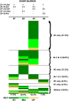
Fig 2. Behavioral responses of V-S (H. virescens donor antennal disc, H. subflexa host) transplant males.
Transplant males exhibited a strong behavioral preference for blends containing Z11-16:Ald, Z9-14:Ald and either Z11-16:OH or Z11-16:OAc (blends 3 and 2 respectively). Over 70% of males that exhibited a behavioral response (N = 188) responded to one or both of these blends. Other males responded to either of the other two blends (#1, H. virescens blend; #4, H. subflexa blend) or to various combinations of the tested mixtures. Source contact (SC) is indicated by dark green, half way to the source (Mid) by a lighter green and upwind flight (UP) by orange. Numbers under each column represent percentage of total responses to that particular odor source (N = 269). Ratios of odorants in each blend are indicated above the four treatments.
Fig 3
Fig 3. Behavioral responses of V-V transplant male controls.
Of 43 males that exhibited positive behavioral responses, 42 responded only to the H. virescens blend (Z11-16:Ald + Z9-14:Ald, 1:0.1). Only a single male responded to the H. zea blend (Z11-16:Ald + Z9-16:Ald, 1:0.1). Source contact (SC) is indicated by dark green, half way to the source (Mid) by a lighter green and upwind flight (UP) by orange. Ratios of odorants in each blend are indicated above the four treatments.
Fig 4
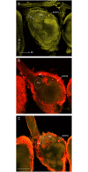
Fig 4. Physiology and morphology of transplant male projection neurons associated with ‘expected’ glomerular targets.
Projection neurons (PNs) that responded specifically to stimulation with either Z11-16:Ald (A, B) or Z9-14:Ald (C, D). Both PNs had cell bodies located in the medial cell body cluster. Images (B, D) represent projections of a series of confocal images. Approximate locations of glomerular boundaries, which tend to be blurred in multiple image projections, are indicted by a dotted line. (A) Physiological profile of a neuron tuned to Z11-16:Ald. (B) A single neuron was stained in this preparation and shown to have a dendritic arborization restricted to the cumulus (Cu). The dorso-medial glomerulus (DM) is visible in this projection but the antero-medial and ventro-medial glomeruli (AM and VM respectively) have receded at this depth in the antennal lobe. (C) Physiological profile of a neuron tuned specifically to Z9-14:Ald. (D) Two PNs were stained in this preparation. One had a dendritic arbor restricted the DM glomerulus, consistent with Z9-14:Ald PNs in normal H. virescens males. A second neuron was localized to an ordinary glomerulus (og). In this more anterior image both AM and VM glomeruli are visible. Scale bar = 100μm (B, D); D, dorsal, M, medial.
Fig 5
Fig 5. Physiology of the four projection neurons that accompany stained neurons in Fig 6.
(A) Neuron with specific responses to Z11-16:Ald (morphology in Fig 6A). (B) From the same preparation, a PN that responded only to antennal stimulation with Z9-14:Ald (morphology in Fig 6B). (C) A PN that responded similarly to stimulation with Z9-14:Ald (morphology in Fig 6C). (D) PN responsive specifically to Z11-16:OH (morphology in Fig 6D).
Fig 6
Fig 6. Projection neurons connected with the antero-medial (AM) and ventro-medial (VM) glomeruli had variable odorant associations in transplant males.
(A) Z11-16:Ald projection neuron (PN) (stained with TMR-dextran) with a dendritic arbor restricted to the cumulus. (B) In the same preparation a Z9-14:Ald PN (stained with a different fluorescent marker–Lucifer yellow) exhibited a dense dendritic arbor restricted to the VM glomerulus. Note that some branches of this PN passage through the AM glomerulus but it was unclear if synaptic contacts were made within this glomerulus. (C) A Z9-14:Ald-specific PN associated with the AM glomerulus. In both B and C, dendritic arbors would be expected in the dorso-medial glomerulus (DM). (D) A Z11-16:OH PN exhibited a dense dendritic arbor in the VM glomerulus. Note that a second PN, associated with the cumulus, was also stained in this preparation. This PN responded to Z11-16:Ald. Scale bar = 100μm; D, dorsal, M, medial.
Fig 7
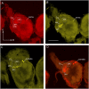
Fig 7. Projection neurons recorded from V-V control transplant males show a normal pattern of arborization in the MGC.
(A) Medial cell body PN (MCB-PN) with an arbor restricted to the cumulus (Cu) responded to Z11-16:Ald. (B) MCB-PN with a dense dendritic arborization restricted to the dorso-medial glomerulus (DM) responded to stimulation with Z9-14:Ald. (C) In this preparation, two MCB-PNs with arbors restricted to the ventro-medial glomerulus (VM) and the cumulus were stained. The PN that responded to Z11-16:OH was associated with the VM glomerular location. Scale bar = 100μm; D, dorsal, M, medial.
Fig 8
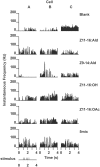
Fig 8. Physiological response profiles of the three projection neurons shown in Fig 7.
Responses are shown as instantaneous frequency plots where responses to stimulation with 5 brief pulses of a specific odorant (or a mixture containing the odorant) result in volleys of action potentials seen as distinct peaks above background. (A) PN specifically responsive to stimulation with Z11-16:Ald or mixtures containing that odorant (morphology shown in Fig 7A). (B) PN specifically responsive to Z9-14:Ald or mixtures with this odorant (morphology shown in Fig 7B). Note strong hyperpolarizing responses to pulses of Z11-16:Ald and Z11-16:OH. (C) PN responsive to Z11-16:OH and blends that contained this odorant (morphology shown in Fig 7C).
Fig 9
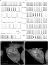
Fig 9. Response profiles and glomerular targets of olfactory receptor neurons (ORNs) from long trichoid sensilla on the antennae of transplant males.
Recordings from V-S transplant males confirmed the presence of sensillar types that are typical for a normal H. virescens antenna. (A-C) Recording from a A-type sensillum housing a Z11-16:Ald ORN (A). Staining revealed projections to the cumulus and PCx1 glomeruli (B, C). (D-F) Recording from a B-type sensillum containing a Z9-14:Ald ORN (D). Staining revealed projections from this sensillum to the dorso-medial glomerulus (DM) and a posterior complex glomerulus (PCx) (E, F). (G-H) Recording from a C-type sensillum containing ORNs responsive to Z11-16:OAc and Z11-16:OH (G). Staining revealed a projection to the antero-medial glomerulus (AM) in this preparation–an observation that is consistent with the normal arborization pattern of ORNs associated with this sensillum type (H). The other paired ORN in the C-type sensillum typically projects to the ventro-medial glomerulus (VM) (but stains of axonal terminals in VM were not seen clearly in this preparation). Some silver-intensified cobalt stains of axon terminals are indicated by small white arrowheads in each section. Scale bar = 100μm (for histological images: B, C, E, F, H); D, dorsal, M, medial–axes the same for each histological image.

Similar articles

Cited by

  • Evolution of olfactory circuits in insects.
    Zhao Z, McBride CS. Zhao Z, et al. J Comp Physiol A Neuroethol Sens Neural Behav Physiol. 2020 May;206(3):353-367. doi: 10.1007/s00359-020-01399-6. Epub 2020 Jan 27. J Comp Physiol A Neuroethol Sens Neural Behav Physiol. 2020. PMID: 31984441 Free PMC article. Review.

References

    1. de Bruyne M, Baker TC (2008) Odor detection in insects: volatile codes. J Chem Ecol 34:882–897. 10.1007/s10886-008-9485-4 - DOI - PubMed
    1. Lei H, Vickers N (2008) Central processing of natural odor mixtures in insects. J Chem Ecol 34: 915–927. 10.1007/s10886-008-9487-2 - DOI - PubMed
    1. Galizia CG, Szyszka P (2008) Olfactory coding in the insect brain: molecular receptive ranges, spatial and temporal coding. Ent Exp Appl 128: 81–92.
    1. Fitt GP (1989) The ecology of Heliothis species in relation to agroecosystems. Ann Rev Entomol 34: 17–52.
    1. Mitter C, Poole RW, Matthews M (1993) Biosystematics of the Heliothinae (Lepidoptera: Noctuidae). Ann Rev Entomol 38: 207–225.

Publication types

LinkOut - more resources