

Preliminary Experience with a New Multidirectional Videoendoscope for Neuroendoscopic Surgical Procedures
- PMID: 26816293
- PMCID: PMC4729436
- DOI: 10.1371/journal.pone.0147524
Preliminary Experience with a New Multidirectional Videoendoscope for Neuroendoscopic Surgical Procedures
Abstract
Purpose: We assessed the applicability of a new multidirectional videoendoscope (digiCAMeleon, Karl Storz GmbH, Tuttlingen, Germany) in various neuroendoscopic procedures.
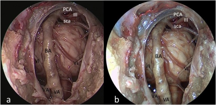
Methods: A 4-mm-diameter rigid videoendoscope (digiCAMeleon, Karl Storz GmbH, Tuttlingen, Germany) with 1 sensor and an internal LED light source was tested. The device offers a resolution of 1920 x 1080 pixels and weighs ≈ 215 g. The prototype was tested on three cadaveric heads using three different approaches: a) endoscopic endonasal transsphenoidal; b) frontal transcortical intraventricular; c) supraorbital.
Results: We identified several major benefits of the integrated system as applied to endoscopic endonasal, transcortical intraventricular, and endoscopic supraorbital keyhole approaches. These included improved maneuverability of the scope on account of reduced bulk and integration of the camera and fiberoptic light components, a variable angle of view from 0-70 degrees, and a novel feature that can be activated to maintain orientation of the surgical horizon. Our preliminary report highlights the potential for handling the videoendoscope in one hand, as one would a microsurgical instrument. The videoendoscope harbors all its electronic and lighting data into a unique and thin cable, thus resembling a modern "all-in-one" computer technology. Because of its reduced weight and ergonomic shape, controlling its movements is very easy and comfortable, even in the microsurgical environment. Furthermore, the videoendoscope offers the unique feature of orienting the horizon of vision, thanks to the possibility of offering angled views while working; this helps the surgeons to stay oriented with direct visualization and improved control of the instruments over a specific area of interest.
Conclusions: The videoendoscope prototype represents an HD-image quality versatile tool in a neurosurgical environment, thanks to its reduced weight and dimensions; in these preliminary simulations, we have identified optimized visibility and maneuverability as major benefits of this novel surgical adjunct.
Conflict of interest statement
Figures


Similar articles
-
Actual state of EndActive ventricular endoscopy.Childs Nerv Syst. 2012 Jan;28(1):87-91. doi: 10.1007/s00381-011-1537-3. Epub 2011 Aug 18. Childs Nerv Syst. 2012. PMID: 21850468
-
Broadening horizons of neuroendoscopy with a variable-view rigid endoscope: an anatomical study.Eur J Surg Oncol. 2010 Feb;36(2):195-200. doi: 10.1016/j.ejso.2009.07.185. Epub 2009 Aug 27. Eur J Surg Oncol. 2010. PMID: 19716259
-
Developments in neuroendoscopy: trial of a miniature rigid endoscope with a multidirectional steerable tip camera in the anatomical lab.Neurosurg Rev. 2012 Jan;35(1):45-50; discussion 50-1. doi: 10.1007/s10143-011-0341-6. Epub 2011 Jul 30. Neurosurg Rev. 2012. PMID: 21805114
-
General principles and intraventricular neuroendoscopy: endoscopic techniques.World Neurosurg. 2013 Feb;79(2 Suppl):S14.e23-8. doi: 10.1016/j.wneu.2012.02.031. Epub 2012 Feb 10. World Neurosurg. 2013. PMID: 22381832 Review.
-
Instrumentation: endoscopes and equipment.World Neurosurg. 2013 Feb;79(2 Suppl):S14.e11-21. doi: 10.1016/j.wneu.2012.02.032. Epub 2012 Feb 10. World Neurosurg. 2013. PMID: 22381831 Review.
Cited by
-
Application of the Endoscopic Micro-Inspection Tool QEVO® in the Surgical Treatment of Anterior Circulation Aneurysms-A Technical Note and Case Series.Front Surg. 2020 Nov 24;7:602080. doi: 10.3389/fsurg.2020.602080. eCollection 2020. Front Surg. 2020. PMID: 33330612 Free PMC article.
References
Publication types
MeSH terms
LinkOut - more resources
Full Text Sources
Other Literature Sources
