Case Reports
. 2016 Jan;31(1):60-4.
doi: 10.5001/omj.2016.11.

Malignant Gastric Glomus Tumor: A Case Report and Literature Review of a Rare Entity

Affiliations
Case Reports

Malignant Gastric Glomus Tumor: A Case Report and Literature Review of a Rare Entity

Shaesta Zaidi et al. Oman Med J. 2016 Jan.

Abstract

A glomus tumor is a mesenchymal neoplasm that usually develops in the peripheral soft tissue, especially in the distal part of the extremities. The subungual zones of the fingers and toes are the most frequent sites of observation. The majority of glomus tumors are entirely benign, and the malignant counterparts are very rare, especially those arising in the visceral organs. We report a case of an extremely rare malignant glomus tumor arising in the stomach of a 53-year-old female admitted to the King Khalid University Hospital, Saudi Arabia. The patient reported a four-month history of pain and fullness in the left hypochondrium. She underwent laparotomy and resection of the gastric mass. The mass was analysed by histopathology. Based on the pathological findings of large tumor size, nuclear atypia, increased mitotic rate, atypical mitosis, the presence of necrosis, and characteristic immunohistochemistry the diagnosis of malignant glomus tumor was rendered. Ultrastructural study confirmed the diagnosis. The patient is well and continues regular follow-up.

Keywords: Glomus Tumor; Stomach; Tumors.


Figures

Figure 1
Figure 1
Computed tomography scan of the abdomen showing a well-defined cystic mass in the subdiaphragmatic region in close contact with the lateral wall of the stomach, and with thick internal septations.
Figure 2
Figure 2
Gross cut section of the mass attached to the wall of the stomach. The mass was multicystic and measured 10 × 9 × 7.5 cm.
Figure 3
Figure 3
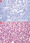
(a) Hematoxylin and eosin staining. Branching vascular channels separated by stroma containing glomus cells in nests, aggregates. The tumor cells are uniform, round, sharply demarcated, with distinct cell borders, arranged around prominent, dilated vascular channels, magnification = 200 ×. (b) Areas showing overall maintenance of architecture, but with malignant appearing round cells showing increased cellularity, pleomorphism, prominent nucleoli, and atypical mitosis, magnification = 400 ×.
Figure 4
Figure 4
(a) Proliferative index (Ki-67) is approximately 15% in the cellular areas, magnification = 200 ×. Cells stained positively for (b) alpha smooth muscle actin, (c) h-caldesmon, (d) vimentin, (e) collagen type IV, and (f) synaptophysin. Magnification of b, c, d, f = 400 ×. Magnification of e = 200 ×.
Figure 5
Figure 5
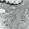
Electron microscopy showing tumor cells invaded by dense basal lamina and perinuclear myofilaments. Magnification = 12,000 ×.

References

    1. Herawi M, Parwani AV, Edlow D, Smolev JK, Epstein JI. Glomus tumor of renal pelvis: a case report and review of the literature. Hum Pathol 2005. Mar;36(3):299-302. 10.1016/j.humpath.2004.10.005 - DOI - PubMed
    1. Miettinen M, Paal E, Lasota J, Sobin LH. Gastrointestinal glomus tumors: a clinicopathologic, immunohistochemical, and molecular genetic study of 32 cases. Am J Surg Pathol 2002. Mar;26(3):301-311. 10.1097/00000478-200203000-00003 - DOI - PubMed
    1. Silver SA, Tavassoli FA. Glomus tumor arising in a mature teratoma of the ovary: report of a case simulating a metastasis from cervical squamous carcinoma. Arch Pathol Lab Med 2000. Sep;124(9):1373-1375. - PubMed
    1. Folpe AL, Fanburg-Smith JC, Miettinen M, Weiss SW. Atypical and malignant glomus tumors: analysis of 52 cases, with a proposal for the reclassification of glomus tumors. Am J Surg Pathol 2001. Jan;25(1):1-12. 10.1097/00000478-200101000-00001 - DOI - PubMed
    1. Kang G, Park HJ, Kim JY, Choi D, Min BH, Lee JH, et al. Glomus tumor of the stomach: a clinicopathologic analysis of 10 cases and review of the literature. Gut Liver 2012. Jan;6(1):52-57. 10.5009/gnl.2012.6.1.52 - DOI - PMC - PubMed

Publication types

LinkOut - more resources