The effect of bacterial cellulose membrane compared with collagen membrane on guided bone regeneration
- PMID: 26816579
- PMCID: PMC4722153
- DOI: 10.4047/jap.2015.7.6.484
The effect of bacterial cellulose membrane compared with collagen membrane on guided bone regeneration
Abstract
Purpose: This study was to evaluate the effects of bacterial cellulose (BC) membranes as a barrier membrane on guided bone regeneration (GBR) in comparison with those of the resorbable collagen membranes.
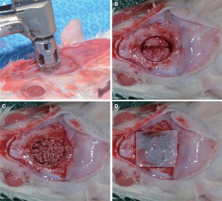
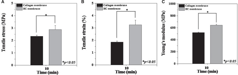
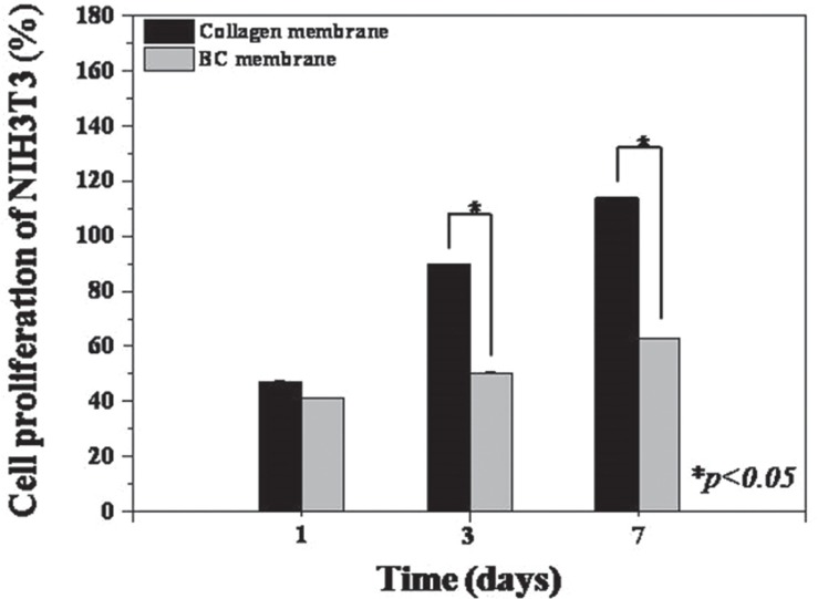
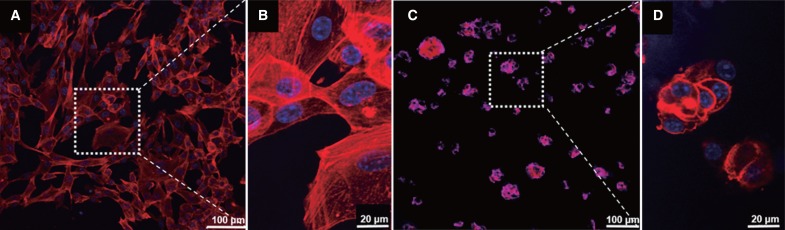
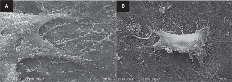
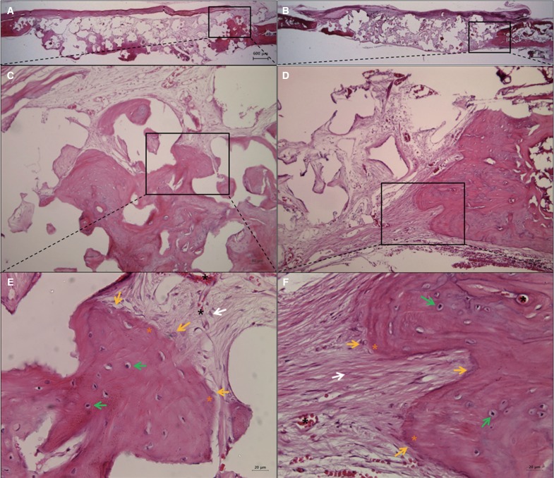
Materials and methods: BC membranes were fabricated using biomimetic technology. Surface properties were analyzed, Mechanical properties were measured, in vitro cell proliferation test were performed with NIH3T3 cells and in vivo study were performed with rat calvarial defect and histomorphometric analysis was done. The Mann-Whitney U test and the Wilcoxon signed rank test was used (α<.05).
Results: BC membrane showed significantly higher mechanical properties such as wet tensile strength than collagen membrane and represented a three-dimensional multilayered structure cross-linked by nano-fibers with 60 % porosity. In vitro study, cell adhesion and proliferation were observed on BC membrane. However, morphology of the cells was found to be less differentiated, and the cell proliferation rate was lower than those of the cells on collagen membrane. In vivo study, the grafted BC membrane did not induce inflammatory response, and maintained adequate space for bone regeneration. An amount of new bone formation in defect region loaded with BC membrane was significantly similar to that of collagen membrane application.
Conclusion: BC membrane has potential to be used as a barrier membrane, and efficacy of the membrane on GBR is comparable to that of collagen membrane.
Keywords: Bacterial cellulose membrane; Collagen membrane; Guided bone regeneration; Rat calvarial defect.
Figures


Similar articles
-
The Efficacy of Electron Beam Irradiated Bacterial Cellulose Membranes as Compared with Collagen Membranes on Guided Bone Regeneration in Peri-Implant Bone Defects.Materials (Basel). 2017 Sep 1;10(9):1018. doi: 10.3390/ma10091018. Materials (Basel). 2017. PMID: 28862689 Free PMC article.
-
Preparation and Characterization of Resorbable Bacterial Cellulose Membranes Treated by Electron Beam Irradiation for Guided Bone Regeneration.Int J Mol Sci. 2017 Oct 25;18(11):2236. doi: 10.3390/ijms18112236. Int J Mol Sci. 2017. PMID: 29068426 Free PMC article.
-
Evaluation of nanohydroxyapaptite (nano-HA) coated epigallocatechin-3-gallate (EGCG) cross-linked collagen membranes.Mater Sci Eng C Mater Biol Appl. 2017 Sep 1;78:258-264. doi: 10.1016/j.msec.2017.04.069. Epub 2017 Apr 18. Mater Sci Eng C Mater Biol Appl. 2017. PMID: 28575983
-
Collagen based barrier membranes for periodontal guided bone regeneration applications.Odontology. 2017 Jan;105(1):1-12. doi: 10.1007/s10266-016-0267-0. Epub 2016 Sep 9. Odontology. 2017. PMID: 27613193 Review.
-
[Research progress on the modification of guided bone regeneration membranes].Hua Xi Kou Qiang Yi Xue Za Zhi. 2019 Jun 1;37(3):325-329. doi: 10.7518/hxkq.2019.03.019. Hua Xi Kou Qiang Yi Xue Za Zhi. 2019. PMID: 31218871 Free PMC article. Review. Chinese.
Cited by
-
De novo strategy with engineering a multifunctional bacterial cellulose-based dressing for rapid healing of infected wounds.Bioact Mater. 2021 Nov 3;13:212-222. doi: 10.1016/j.bioactmat.2021.10.043. eCollection 2022 Jul. Bioact Mater. 2021. PMID: 35224303 Free PMC article.
-
Bacterial Cellulose-Modified Polyhydroxyalkanoates Scaffolds Promotes Bone Formation in Critical Size Calvarial Defects in Mice.Materials (Basel). 2020 Mar 21;13(6):1433. doi: 10.3390/ma13061433. Materials (Basel). 2020. PMID: 32245214 Free PMC article.
-
Opportunities of Bacterial Cellulose to Treat Epithelial Tissues.Curr Drug Targets. 2019;20(8):808-822. doi: 10.2174/1389450120666181129092144. Curr Drug Targets. 2019. PMID: 30488795 Free PMC article. Review.
-
Perspective Applications and Associated Challenges of Using Nanocellulose in Treating Bone-Related Diseases.Front Bioeng Biotechnol. 2021 May 7;9:616555. doi: 10.3389/fbioe.2021.616555. eCollection 2021. Front Bioeng Biotechnol. 2021. PMID: 34026739 Free PMC article. Review.
-
Is the Bacterial Cellulose Membrane Feasible for Osteopromotive Property?Membranes (Basel). 2020 Sep 12;10(9):230. doi: 10.3390/membranes10090230. Membranes (Basel). 2020. PMID: 32932731 Free PMC article.
References
-
- Brånemark PI, Zarb GA, Albrektsson T. Tissue-integrated prostheses: Osseointegration in clinical dentistry. Chicago: Quintessence; 1985. pp. 199–209.
-
- Misch CM. Comparison of intraoral donor sites for onlay grafting prior to implant placement. Int J Oral Maxillofac Implants. 1997;12:767–776. - PubMed
-
- Oda T, Sawaki Y, Ueda M. Experimental alveolar ridge augmentation by distraction osteogenesis using a simple device that permits secondary implant placement. Int J Oral Maxillofac Implants. 2000;15:95–102. - PubMed
-
- Hämmerle CH, Karring T. Guided bone regeneration at oral implant sites. Periodontol 2000. 1998;17:151–175. - PubMed
-
- Ashley FL, Stone RS, Alonsoartieda M, Syverud JM, Edwards JW, Sloan RF, Mooney SA. Experimental and clinical studies on the application of monomolecular cellulose filter tubes to create artificial tendon sheaths in digits. Plast Reconstr Surg Transplant Bull. 1959;23:526–534. - PubMed
LinkOut - more resources
Full Text Sources
Other Literature Sources