Review
. 2014 Nov;1(1):91-102.
doi: 10.1093/rb/rbu011. Epub 2014 Oct 20.

Recent development of temperature-responsive surfaces and their application for cell sheet engineering

Affiliations
Review

Recent development of temperature-responsive surfaces and their application for cell sheet engineering

Zhonglan Tang et al. Regen Biomater. 2014 Nov.

Abstract

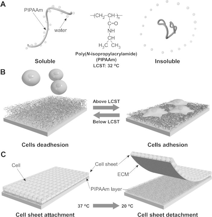
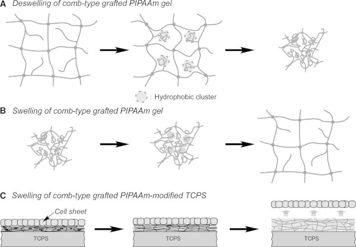
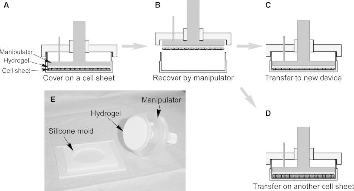
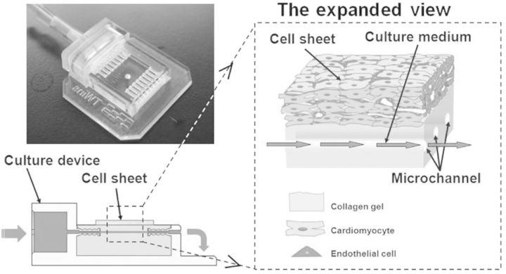
Cell sheet engineering, which fabricates sheet-like tissues without biodegradable scaffolds, has been proposed as a novel approach for tissue engineering. Cells have been cultured and proliferate to confluence on a temperature-responsive cell culture surface at 37°C. By decreasing temperature to 20°C, an intact cell sheet can be harvested from the culture surface without enzymatic treatment. This new approach enables cells to keep their cell-cell junction, cell surface proteins and extracellular matrix. Therefore, recovered cell sheet can be easily not only transplanted to host tissue, but also constructed a three-dimensional (3D) tissue by layering cell sheets. Moreover, cell sheet manipulation technology and bioreactor have been combined with the cell sheet technology to fabricate a complex and functional 3D tissue in vitro. So far, cell sheet technology has been applied in regenerative medicine for several tissues, and a number of clinical studies have been performed. In this review, recent advances in the preparation of temperature-responsive cell culture surface, the fabrication of organ-like tissue and the clinical application of cell sheet engineering are summarized and discussed.

Keywords: cell sheet; cell sheet engineering; poly(N-isoproplyacrylamide); temperature-responsive cell culture surface.


Figures

Figure 1.
Figure 1.
Schematic illustration of the temperature-responsive character change of PIPAAm in aqueous solution (A) and on substrate (B). Attaching cells can proliferate to confluence, and detach from PIPAAm-grafted substrate with ECM, when decreasing temperature from 37°C to 20°C (C).
Figure 2.
Figure 2.
(A) Schematic illustration of the fabricating a micropatterned temperature-responsive cell culture surface. (B) Schematic illustration of the modified LCD projector for maskless photolithography. (C) Phase contrast microscopic photographs of human umbilical vein endothelial cells HUVECs on 20 µm-wide adhesive PIPAAm-grafted domains at 37°C. Scale bar: 100 µm. (Reprinted with permission from Ref. [75] © 2007 Elsevier.)
Figure 3.
Figure 3.
(A) Schematic illustration of the fabrication of micropatterned temperature-responsive PIPAAm/PIPAAm-b-PAcMo patterned brush surface via a two-step RAFT polymerization. PAcMo have been grafted from the terminal of PIPAAm chains via reactive DTB groups. (B) Phase contrast image shows that NHDFs only adhere on 100 µm-wide PIPAAm regions of PIPAAm/PIPAAm-b-PAcMo patterned surface after 24 h culture. (C) Phase contrast image and (D) fluorescence image show that NHDF sheet is formed on a 50-µm-wide PIPAAm/PIPAAm-b-PAcMo patterned surface after a 5-day culture. (D) Actin and nuclei of NHDFs are stained with AlexaFluor568-phalloidin (red) and Hoechst 33258 (blue), respectively. Scale bars: 100 µm. (Reprinted with permission from Ref. [78] © 2011 the American Chemical Society.)
Figure 4.
Figure 4.
Schematic illustration of water penetration of (A) PIPAAm-grafted TCPS, (B) PIPAAm-grafted PM and (C) P(IPAAm-co-PEG)-grafted PM during cell sheet detachment.
Figure 5.
Figure 5.
Schematic illustration of (A) deswelling and (B) swelling of comb-type-grafted PIPAAm gel and (C) the swelling of cell culture surface grafting with comb-type-grafted PIPAAm. (Reprinted with permission from Ref. [83] © 2010 Elsevier.)
Figure 6.
Figure 6.
Schematic illustration of the cell sheet manipulator for transferring cell sheet and stacking cell sheet. (A) Cell sheet has been covered with a hydrogel-coated manipulator and incubated for 20 min at 20°C. (B) Cell sheet has been recovered by the manipulator. (C) The harvested cell sheet has been transferred to a new cell culture dish. (D) The recovered cell sheet has been stacked on another cell sheet. (E) Macroscopic view of a cell sheet manipulation device.
Figure 7.
Figure 7.
Schematic illustration of the cell sheet culture device with tissue surrogates. The left picture shows a photograph of the culture device.

Similar articles

Cited by

References

    1. Atala A, Lanza R, Thomson JA, et al. Principles of Regenerative Medicine, 2nd edn New York: Academic Press, 2011.
    1. Mozid AM, Arnous S, Sammut EC, et al. Stem cell therapy for heart diseases. Br Med Bull 2001;98:143–59. - PubMed
    1. Pereira DR, Silva-Correia J, Oliveira JM, et al. Hydrogels in acellular and cellular strategies for intervertebral disc regeneration. J Tissue Eng Regen Med 2013;7:85–98. - PubMed
    1. Aguirre A, Sancho-Martinez I, Izpisua Belmonte JC. Reprogramming toward heart regeneration: stem cells and beyond. Cell Stem Cell 2013;12:275–84. - PubMed
    1. Mabed M, Shahin M. Mesenchymal stem cell-based therapy for the treatment of type 1 diabetes mellitus. Curr Stem Cell Res Ther 2012;7:179–90. - PubMed