. 2015 Mar;2(1):21-9.
doi: 10.1093/rb/rbu018. Epub 2015 Feb 9.

Selectivity of biopolymer membranes using HepG2 cells

Affiliations

Selectivity of biopolymer membranes using HepG2 cells

Dongyuan Lü et al. Regen Biomater. 2015 Mar.

Abstract

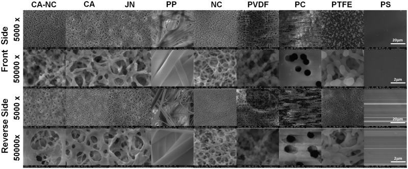
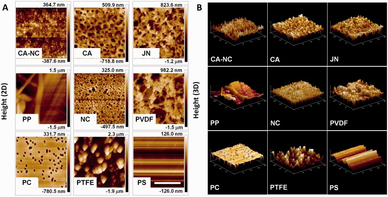
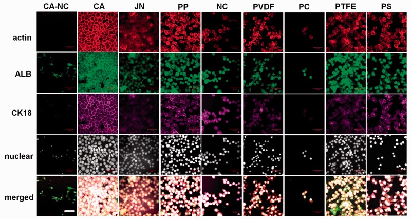
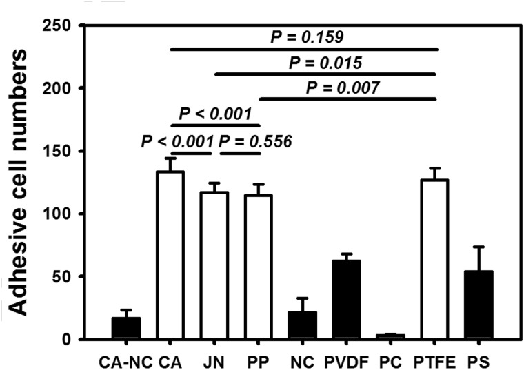
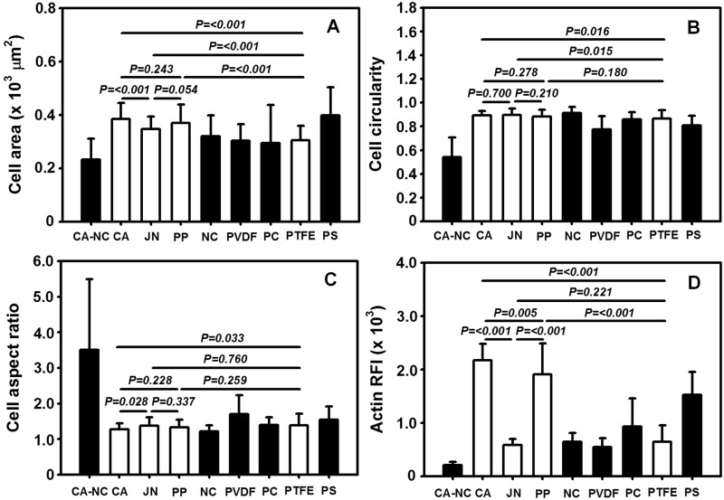
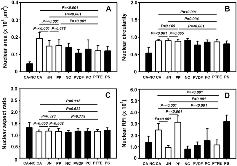
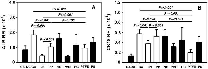
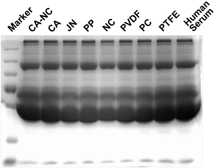
Bioartificial liver (BAL) system has emerged as an alternative treatment to bridge acute liver failure to either liver transplantation or liver regeneration. One of the main reasons that the efficacy of the current BAL systems was not convincing in clinical trials is attributed to the lack of friendly interface between the membrane and the hepatocytes in liver bioreactor, the core unit of BAL system. Here, we systematically compared the biological responses of hepatosarcoma HepG2 cells seeded on eight, commercially available biocompatible membranes made of acetyl cellulose-nitrocellulose mixed cellulose (CA-NC), acetyl cellulose (CA), nylon (JN), polypropylene (PP), nitrocellulose (NC), polyvinylidene fluoride (PVDF), polycarbonate (PC) and polytetrafluoroethylene (PTFE). Physicochemical analysis and mechanical tests indicated that CA, JN and PP membranes yield high adhesivity and reasonable compressive and/or tensile features with friendly surface topography for cell seeding. Cells prefer to adhere on CA, JN, PP or PTFE membranes with high proliferation rate in spheriod-like shape. Actin, albumin and cytokeratin 18 expressions are favorable for cells on CA or PP membrane, whereas protein filtration is consistent among all the eight membranes. These results further the understandings of cell growth, morphology and spreading, as well as protein filtration on distinct membranes in designing a liver bioreactor.

Keywords: HepG2; bioartificial liver; biocompatible membrane.


Figures

Figure 1.
Figure 1.
membrane structures via SEM images of eight membranes or control PS slide. Front (rows first and second) or reverse (rows third ans fourth) side of membrane or slide was viewed at low (×5000, rows first and third) or high (×50 000, rows second and fourth) magnification.
Figure 2.
Figure 2.
surface microtography via AFM images of eight membranes or control PS plate. Front side of membrane or plate was viewed from a 2D (A) or 3D (B) demonstration. Bar = 10 µm. Scale of height in 2D images was presented on the right scale bar of each panel in A.
Figure 3.
Figure 3.
hepatic biomarker expression of HepG2 cells grown on the eight membranes or PS substrate. Data were presented as the fluorescent staining of actin (first row), ALB (second row) or CK18 (third row) proteins and of nucleus presentation (fourth row) or merged co-localization (fifth row) at t = 24 h at × 63 (Bar = 50 µm).
Figure 4.
Figure 4.
determination of adhesion capacity of HepG2 cells via number of adhered HepG2 cells on the eight membranes or PS substrate at t = 24 h. Totally 10 frame data were obtained from the repeats in triplets and presented as the mean ± standard deviation (SD) per frame.
Figure 5.
Figure 5.
morphology and spreading of adhered HepG2 cells on eight membranes or PS substrate. Plotted are cellular projected area (A), circularity (B), aspect ratio (C) as well as relative fluorescence intensity of actin (D) at t = 24 h. Totally 10 frame data were obtained from the repeats in triplets and presented as the mean ± SD.
Figure 6.
Figure 6.
nucleus morphology and spreading of HepG2 cells on eight membranes or PS substrate. Plotted are nuclear projected area (A), circularity (B), aspect ratio (C) as well as relative fluorescence intensity of nucleus (D) at t = 24 h. Totally 10 frame data were obtained from the repeats in triplets and presented as the mean ± SD.
Figure 7.
Figure 7.
comparison of ALB (A) and CK18 (B) expressions on distinct membranes at t = 24 h. Data were obtained from the repeats in triplets and presented as the mean ± SD.
Figure 8.
Figure 8.
human serum filtration through distinct membranes. Filtrated components were collected and tested using SDS–PAGE electrophoresis assay.

Similar articles

Cited by

References

    1. Gebhardt R. Metabolic zonation of the liver: regulation and implications for liver function. Pharmacol Ther 1992;53:275–354. - PubMed
    1. Farmer D, Anselmo D, Ghobrial R, et al. Liver transplantation for fulminant hepatic failure: experience with more than 200 patients over a 17-year period. Ann Surg 2003;237:666–76. - PMC - PubMed
    1. Park J, Lee D. Bioartificial liver systems: current status and future perspective. J Biosci Bioeng 2005;99:311–9. - PubMed
    1. Opolon P. High permeability membrane hemodialysis and hemofiltration in acute hepatic coma: experimental and clinical results. Artif Organs 1979;3:354–60. - PubMed
    1. Knell A, Dukes D. Dialysis procedures in acute liver failure. Lancet 1976;2:402–3. - PubMed