

Application of Raman spectroscopy in Andrology: non-invasive analysis of tissue and single cell
- PMID: 26816760
- PMCID: PMC4708293
- DOI: 10.3978/j.issn.2223-4683.2014.03.01
Application of Raman spectroscopy in Andrology: non-invasive analysis of tissue and single cell
Abstract
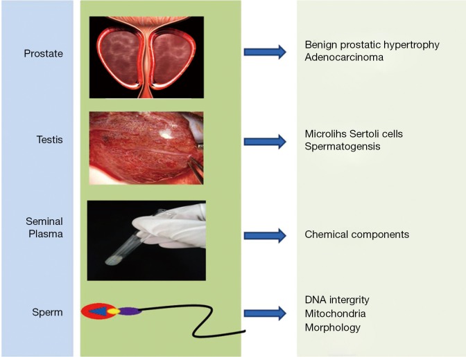
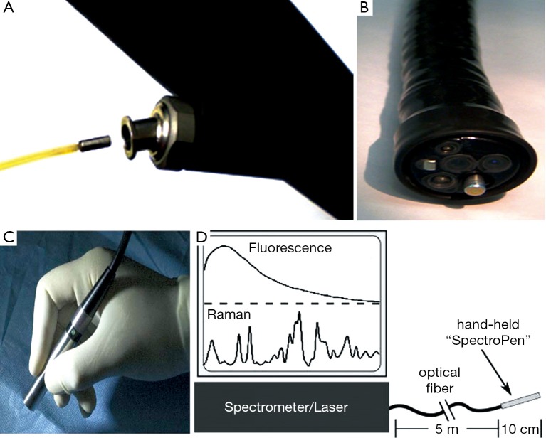
As a fast, label-free and non-invasive detection method, Raman spectroscopy has been widely used for the interrogation of biological tissues, any alterations of molecular structure and chemical components during pathological processes would be identified and revealed via the differences on Raman spectrum. In clinics, the Raman spectroscopy has great potentials to provide real-time scanning of living tissues and fast diagnosis of diseases, just like discrimination of various carcinomas. A portable Raman spectroscopy which combined Raman system with an optical fiber probe has also been developed and proved to be able to provide intraoperative assistance in both human study and animal models. In Andrology, interests in Raman spectroscopy had just emerged. In this review, we summarized the progress about the utility of Raman spectroscopy in Andrology, the literatures were gathered from PubMed and Ovid database using MeSH terms associated with prostate, testis, seminal plasma and single sperm cell. We also highlighted the serious challenges as to the final clinical application of Raman technique. In conclusion, research in Raman spectroscopy may herald a new era for Andrology.
Keywords: Andrology; Raman spectroscopy; clinical application; non-invasive.
Conflict of interest statement
Figures
Similar articles
-
Application and Progress of Raman Spectroscopy in Male Reproductive System.Front Cell Dev Biol. 2022 Jan 12;9:823546. doi: 10.3389/fcell.2021.823546. eCollection 2021. Front Cell Dev Biol. 2022. PMID: 35096844 Free PMC article. Review.
-
Clinical and investigative applications of Raman spectroscopy in Urology and Andrology.Transl Androl Urol. 2014 Mar;3(1):84-8. doi: 10.3978/j.issn.2223-4683.2014.01.02. Transl Androl Urol. 2014. PMID: 26816755 Free PMC article. Review.
-
Raman spectroscopy and its urological applications.Indian J Urol. 2008 Oct;24(4):444-50. doi: 10.4103/0970-1591.39550. Indian J Urol. 2008. PMID: 19468494 Free PMC article.
-
Label-free Molecular Imaging and Analysis by Raman Spectroscopy.Acta Histochem Cytochem. 2018 Jun 26;51(3):101-110. doi: 10.1267/ahc.18019. Epub 2018 Jun 20. Acta Histochem Cytochem. 2018. PMID: 30083018 Free PMC article.
-
Portable Sequentially Shifted Excitation Raman spectroscopy as an innovative tool for in situ chemical interrogation of painted surfaces.Analyst. 2016 Aug 7;141(15):4599-607. doi: 10.1039/c6an00753h. Epub 2016 Jun 8. Analyst. 2016. PMID: 27273377
Cited by
-
Spectral features of nuclear DNA in human sperm assessed by Raman Microspectroscopy: Effects of UV-irradiation and hydration.PLoS One. 2018 Nov 20;13(11):e0207786. doi: 10.1371/journal.pone.0207786. eCollection 2018. PLoS One. 2018. PMID: 30458032 Free PMC article.
-
Fast diagnosis of men's fertility using Raman spectroscopy combined with chemometric methods: An experimental study.Int J Reprod Biomed. 2021 Feb 21;19(2):121-128. doi: 10.18502/ijrm.v19i2.8470. eCollection 2021 Feb. Int J Reprod Biomed. 2021. PMID: 33718756 Free PMC article.
-
Decoding the role of cytochrome c in metabolism of human spermatozoa by Raman imaging.Front Cell Dev Biol. 2022 Nov 25;10:983993. doi: 10.3389/fcell.2022.983993. eCollection 2022. Front Cell Dev Biol. 2022. PMID: 36506104 Free PMC article.
-
Non-Invasive Approaches to Epigenetic-Based Sperm Selection.Med Sci Monit. 2017 Sep 29;23:4677-4683. doi: 10.12659/msm.904098. Med Sci Monit. 2017. PMID: 28961228 Free PMC article. Review.
-
Sperm selection in IVF: the long and winding road from bench to bedside.JBRA Assist Reprod. 2020 Jul 14;24(3):332-339. doi: 10.5935/1518-0557.20190081. JBRA Assist Reprod. 2020. PMID: 32155013 Free PMC article. Review.
References
-
- Huang WE, Li M, Jarvis RM, et al. Shining light on the microbial world the application of Raman microspectroscopy. Adv Appl Microbiol 2010;70:153-86. - PubMed
-
- Rao AR, Hanchanale V, Javle P, et al. Spectroscopic view of life and work of the Nobel Laureate Sir C.V. Raman. J Endourol 2007;21:8-11. - PubMed
-
- Hanlon EB, Manoharan R, Koo TW, et al. Prospects for in vivo Raman spectroscopy. Phys Med Biol 2000;45:R1-59. - PubMed
-
- Li M, Xu J, Romero-Gonzalez M, et al. Single cell Raman spectroscopy for cell sorting and imaging. Curr Opin Biotechnol 2012;23:56-63. - PubMed
Publication types
LinkOut - more resources
Full Text Sources
Research Materials