

doi: 10.1038/ncomms10534.
Volta phase plate cryo-EM of the small protein complex Prx3
Affiliations
- PMID: 26817416
- PMCID: PMC4738354
- DOI: 10.1038/ncomms10534
Item in Clipboard
Volta phase plate cryo-EM of the small protein complex Prx3
Nat Commun.
.
Abstract
Cryo-EM of large, macromolecular assemblies has seen a significant increase in the numbers of high-resolution structures since the arrival of direct electron detectors. However, sub-nanometre resolution cryo-EM structures are rare compared with crystal structure depositions, particularly for relatively small particles (<400 kDa). Here we demonstrate the benefits of Volta phase plates for single-particle analysis by time-efficient cryo-EM structure determination of 257 kDa human peroxiredoxin-3 dodecamers at 4.4 Å resolution. The Volta phase plate improves the applicability of cryo-EM for small molecules and accelerates structure determination.
Figures
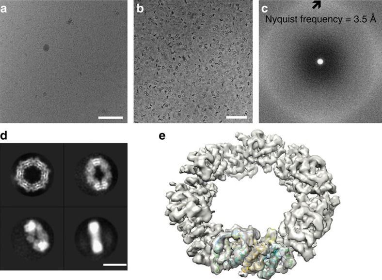
(a) Electron micrograph of frozen-hydrated hPrx3 dodecamers taken without a phase plate at 2.4 μm underfocus. In thick ice, toroids are only readily discernible at high defocus (scale bar, 100 nm). (b) In-focus electron micrograph of hPrx3 particles. VPP images of hPrx3 are high in contrast even in-focus and before motion correction, enabling robust automated particle selection of all views (scale bar, 100 nm). (c) Power spectrum of electron micrograph featuring continuous signal without contrast transfer function (CTF) oscillations across the frequency spectrum. (d) Four representative class averages of hPrx3 featuring random orientations promoted by thick ice (scale bar, 10 nm). (e) Isosurface representation of the reconstructed 3D density map with bovine Prx3 dimer structure docked (PDB 1ZYE (ref. 14)).
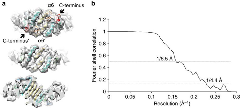
(a) The VPP reconstruction of hPrx3 is consistent with the crystal structure of hPrx2 (PDB 1QMV (ref. 15)), where the dimer features folded C terminus (red; top). The C-terminal region is not present in the crystal structure of the bovine homologue of Prx3 (PDB 1ZYE (ref. 14)) and disappears in our EM map at lower thresholds (middle), suggesting only partial occupancy of the folded C terminus. The obtained resolution is sufficient for resolving individual beta-strands in the density map (bottom). (b) Fourier-shell correlation indicates a resolution of 4.4 Å based on the ‘gold-standard' Fourier shell correlation (FSC)=0.143 criterion.
References
Publication types
MeSH terms
Substances
LinkOut - more resources
Full Text Sources
Other Literature Sources
Miscellaneous
