

CP-CHARM: segmentation-free image classification made accessible
- PMID: 26817459
- PMCID: PMC4729047
- DOI: 10.1186/s12859-016-0895-y
CP-CHARM: segmentation-free image classification made accessible
Abstract
Background: Automated classification using machine learning often relies on features derived from segmenting individual objects, which can be difficult to automate. WND-CHARM is a previously developed classification algorithm in which features are computed on the whole image, thereby avoiding the need for segmentation. The algorithm obtained encouraging results but requires considerable computational expertise to execute. Furthermore, some benchmark sets have been shown to be subject to confounding artifacts that overestimate classification accuracy.
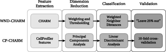
Results: We developed CP-CHARM, a user-friendly image-based classification algorithm inspired by WND-CHARM in (i) its ability to capture a wide variety of morphological aspects of the image, and (ii) the absence of requirement for segmentation. In order to make such an image-based classification method easily accessible to the biological research community, CP-CHARM relies on the widely-used open-source image analysis software CellProfiler for feature extraction. To validate our method, we reproduced WND-CHARM's results and ensured that CP-CHARM obtained comparable performance. We then successfully applied our approach on cell-based assay data and on tissue images. We designed these new training and test sets to reduce the effect of batch-related artifacts.
Conclusions: The proposed method preserves the strengths of WND-CHARM - it extracts a wide variety of morphological features directly on whole images thereby avoiding the need for cell segmentation, but additionally, it makes the methods easily accessible for researchers without computational expertise by implementing them as a CellProfiler pipeline. It has been demonstrated to perform well on a wide range of bioimage classification problems, including on new datasets that have been carefully selected and annotated to minimize batch effects. This provides for the first time a realistic and reliable assessment of the whole image classification strategy.
Figures
Similar articles
-
FogBank: a single cell segmentation across multiple cell lines and image modalities.BMC Bioinformatics. 2014 Dec 30;15(1):431. doi: 10.1186/s12859-014-0431-x. BMC Bioinformatics. 2014. PMID: 25547324 Free PMC article.
-
Phenotype recognition with combined features and random subspace classifier ensemble.BMC Bioinformatics. 2011 Apr 30;12:128. doi: 10.1186/1471-2105-12-128. BMC Bioinformatics. 2011. PMID: 21529372 Free PMC article.
-
Robust and automated three-dimensional segmentation of densely packed cell nuclei in different biological specimens with Lines-of-Sight decomposition.BMC Bioinformatics. 2015 Jun 8;16:187. doi: 10.1186/s12859-015-0617-x. BMC Bioinformatics. 2015. PMID: 26049713 Free PMC article.
-
From pixels to insights: Machine learning and deep learning for bioimage analysis.Bioessays. 2024 Feb;46(2):e2300114. doi: 10.1002/bies.202300114. Epub 2023 Dec 6. Bioessays. 2024. PMID: 38058114 Review.
-
Building cell models and simulations from microscope images.Methods. 2016 Mar 1;96:33-39. doi: 10.1016/j.ymeth.2015.10.011. Epub 2015 Oct 17. Methods. 2016. PMID: 26484733 Free PMC article. Review.
Cited by
-
Automated and Reproducible Detection of Vascular Endothelial Growth Factor (VEGF) in Renal Tissue Sections.J Immunol Res. 2019 Mar 19;2019:7232781. doi: 10.1155/2019/7232781. eCollection 2019. J Immunol Res. 2019. PMID: 31016206 Free PMC article.
-
Rapid 3D phenotypic analysis of neurons and organoids using data-driven cell segmentation-free machine learning.PLoS Comput Biol. 2021 Feb 22;17(2):e1008630. doi: 10.1371/journal.pcbi.1008630. eCollection 2021 Feb. PLoS Comput Biol. 2021. PMID: 33617523 Free PMC article.
-
TDAExplore: Quantitative analysis of fluorescence microscopy images through topology-based machine learning.Patterns (N Y). 2021 Oct 12;2(11):100367. doi: 10.1016/j.patter.2021.100367. eCollection 2021 Nov 12. Patterns (N Y). 2021. PMID: 34820649 Free PMC article.
-
Domain-invariant features for mechanism of action prediction in a multi-cell-line drug screen.Bioinformatics. 2020 Mar 1;36(5):1607-1613. doi: 10.1093/bioinformatics/btz774. Bioinformatics. 2020. PMID: 31608933 Free PMC article.
-
Data-analysis strategies for image-based cell profiling.Nat Methods. 2017 Aug 31;14(9):849-863. doi: 10.1038/nmeth.4397. Nat Methods. 2017. PMID: 28858338 Free PMC article.
References
-
- Rui Y, Huang TS, Chang SF. Image retrieval: Current techniques, promising directions, and open issues. J Vis Commun Image Represent. 1999;10(1):39–62. doi: 10.1006/jvci.1999.0413. - DOI
-
- Huang K, Murphy RF. Proceedings of the IEEE International Symposium on Biomedical Imaging: From Nano to Macro (ISBI’04): April 15–18. Arlington: IEEE; 2004. Automated classification of subcellular patterns in multicell images without segmentation into single cells.
Publication types
MeSH terms
Grants and funding
LinkOut - more resources
Full Text Sources
Other Literature Sources
Miscellaneous
