. 2016 Jan 28:6:19970.
doi: 10.1038/srep19970.

Intracrine Androgens Enhance Decidualization and Modulate Expression of Human Endometrial Receptivity Genes

Affiliations

Intracrine Androgens Enhance Decidualization and Modulate Expression of Human Endometrial Receptivity Genes

Douglas A Gibson et al. Sci Rep. .

Abstract

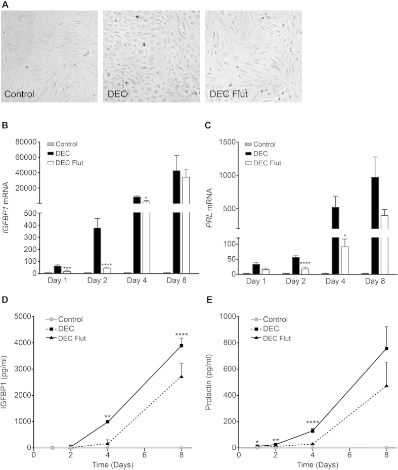
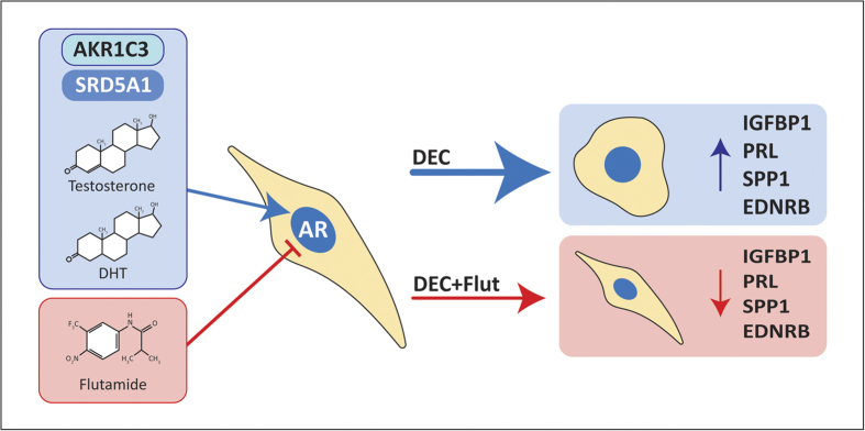
The endometrium is a complex, steroid-dependent tissue that undergoes dynamic cyclical remodelling. Transformation of stromal fibroblasts (ESC) into specialised secretory cells (decidualization) is fundamental to the establishment of a receptive endometrial microenvironment which can support and maintain pregnancy. Androgen receptors (AR) are present in ESC; in other tissues local metabolism of ovarian and adrenal-derived androgens regulate AR-dependent gene expression. We hypothesised that altered expression/activity of androgen biosynthetic enzymes would regulate tissue availability of bioactive androgens and the process of decidualization. Primary human ESC were treated in vitro for 1-8 days with progesterone and cAMP (decidualized) in the presence or absence of the AR antagonist flutamide. Time and treatment-dependent changes in genes essential for a) intra-tissue biosynthesis of androgens (5α-reductase/SRD5A1, aldo-keto reductase family 1 member C3/AKR1C3), b) establishment of endometrial decidualization (IGFBP1, prolactin) and c) endometrial receptivity (SPP1, MAOA, EDNRB) were measured. Decidualization of ESC resulted in significant time-dependent changes in expression of AKR1C3 and SRD5A1 and secretion of T/DHT. Addition of flutamide significantly reduced secretion of IGFBP1 and prolactin and altered the expression of endometrial receptivity markers. Intracrine biosynthesis of endometrial androgens during decidualization may play a key role in endometrial receptivity and offer a novel target for fertility treatment.


Figures

Figure 1
Figure 1. Decidualization is associated with time-dependent changes in expression of AKR1C3 and biosynthesis of testosterone.
The expression of the androgen biosynthetic enzyme AKR1C3, which converts A4 to T, was assessed by qPCR, Western blot and immunocytochemistry in ESC. AKR1C3 was increased following decidualization of ESC. (A) Concentrations of mRNAs encoding AKR1C3 were significantly increased in ESC treated to decidualized (DEC) for 2 and 4 days compared to control (Control, p < 0.0001, n = 8) but by day 8 mRNA expression was unchanged between control and decidualized ESC. (B) Western blot analysis of AKR1C3 expression from homogenates of ESC treated for 4 days revealed a significant increase in AKR1C3 protein in decidualized ESC (n = 4 patients per treatment, p < 0.05). (C) Western blot analysis of AKR1C3 expression from homogenates of ESC treated for 8 days revealed a significant increase in AKR1C3 protein in decidualized ESC (n = 6 patients per treatment, p < 0.05). Representative blots from 4 matched patients are shown (B,C). Loading control b-actin (red, 43 kDa), AKR1C3 (green, 37 kDa). (D) The expression of AKR1C3 was assessed by immunocytochemistry in ESC grown in chamber slides and treated to decidualize for 4 days. AKR1C3 expression was detected in both control and decidualized ESC (AKR1C3; red staining). Staining appeared more intense in decidualized ESC (DEC). Nuclear counterstain SytoxGreen (SYTOX; green staining), scale bar 100 μm. In the absence of primary antibody no staining was detected (inset). (E) Concentrations of T were assessed by ELISA and significant concentrations of T were detected in cell culture supernatants recovered from decidualized ESC at 4 (n = 8, p < 0.01) and 8 days (n = 8, p < 0.01). *p < 0.05, **p < 0.01, ****p < 0.0001.
Figure 2
Figure 2. Decidualization is associated with time-dependent changes in expression of SRD5A1 and biosynthesis of dihydrotestosterone.
The expression of the androgen biosynthetic enzyme SRD5A1, which reduces T to the more potent androgen DHT, was assessed by qPCR, Western blot and immunocytochemistry in ESC. (A) Concentrations of mRNAs encoding SRD5A1 were significantly decreased in ESC treated with DM for 1 and 2 days compared to control (Control, p < 0.01 and p < 0.05 respectively, n = 8) but by day 8 mRNA expression was unchanged between control and decidualized ESC. (B) Western blot analysis of 5α-reductase (SRD5A1) expression from homogenates of ESC treated for 4 days revealed that 5α-reductase protein in was detected in control and decidualized ESC (n = 4 patients per treatment) (C) Western blot analysis of 5α-reductase expression from homogenates of ESC treated for 8 days revealed a significant decrease in 5α-reductase protein in decidualized ESC (n = 7 patients per treatment, p < 0.0001), representative blots from 4 matched patients are shown (B,C). Loading control b-actin (red, 43 kDa), 5α-reductase (green, 100 kDa). (D) The expression of 5α-reductase was assessed by immunocytochemistry in ESC grown in chamber slides and treated to decidualize for 4 days. 5α-reductase expression was detected in both control and decidualized ESC (SRD5A1; Red staining). Staining appeared to be similar in both control and decidualized ESC (DEC). Nuclear counterstain SytoxGreen (SYTOX; green staining), scale bar 100 μm. In the absence of primary antibody no staining was detected (Inset). (E) Concentrations of DHT were assessed by ELISA and significant concentrations of DHT were detected in cell culture supernatants recovered from decidualized ESC at 4 (n = 8, p < 0.0001) and 8 days (n = 8, p < 0.0001). Concentrations of DHT were significantly decreased in day 8 compared to day 4 decidualized ESC (p < 0.001). *p < 0.05, **p < 0.01, ****p < 0.0001.
Figure 3
Figure 3. Blocking local androgen action inhibits decidualization.
To investigate if androgens produced by ESC could affect decidualization, ESC were co-treated with the anti-androgen flutamide and markers of decidual transformation were assessed. (A) Cell morphology was assessed in ESC by phase-contrast microscopy. Control ESC had an elongated fibroblast-like morphology and decidualized (DEC) ESC had a rounded, ‘epithelioid’ morphology. Flutamide treated cells (DEC Flut) were mostly fibroblast-like with few rounded cells. (B) Concentrations of mRNAs encoding IGFBP1 were significantly reduced after 1 (n = 6, p < 0.001), 2 (n = 6, p < 0.0001) and 4 days (n = 6, p < 0.05) of flutamide treatment but were unchanged after 8 days. (C) Concentrations of mRNAs encoding PRL were significantly decreased after 2 (n = 6, p < 0.0001) and 4 days (n = 6, p < 0.05) of flutamide treatment and tended to be lower in flutamide treated ESC after 8 days. (D) Secretion of the decidualization marker IGFBP1 was assessed by ELISA. Secretion of IGFBP1 was significantly reduced by flutamide treatment (DEC Flut). A striking 80% reduction in secretion of IGFBP1 was detected at 4 days (n = 6, p < 0.01) and 30% reduction was detected after 8 days of flutamide treatment (n = 6, p < 0.0001) compared to DEC. (E) Secretion of Prolactin was significantly reduced by flutamide treatment after 1 (n = 6, p < 0.05) and 2 days (n = 6, p < 0.01). A striking 81% reduction in secretion of Prolactin was detected at 4 days (n = 6, p < 0.0001) and secretion of Prolactin tended to be lower in flutamide treated ESC after 8 days compared to DEC ESC (n = 6). *p < 0.05, **p < 0.01, ***p < 0.001, ****p < 0.0001.
Figure 4
Figure 4. The impact of flutamide on expression of putative androgen-regulated receptivity genes.
To investigate if androgens produced by ESC could affect expression of putative androgen-regulated endometrial receptivity genes, ESC were co-treated with the anti-androgen flutamide and the expression of osteopontin (SPP1), monoamine oxidase (MAOA) and endothelin receptor B (EDNRB) were assessed. (A) Concentrations of mRNAs encoding SPP1 were significantly increased in decidualized ESC after 4 (p < 0.05) and 8 days treatment (p < 0.001) and significantly reduced in ESC co-treated with flutamide (n = 8, p < 0.05) at 8 days. (B) Concentrations of secreted SPP1 were detected by ELISA and calculated as fold change relative to decidualized ESC. SPP1 was not detected in supernatants from control cultures (ND). Co-treatment with flutamide significantly reduced secretion of SPP1 (n = 8, p < 0.01) and reduced the relative secretion of SPP1 by ~40%. (C) Concentrations of mRNAs encoding MAOA were significantly increased in decidualized ESC after 1 (p < 0.001), 2 (p < 0.001) and 4 days treatment (p < 0.05). MAOA mRNA expression was significantly increased relative to control in ESC co-treated with flutamide for 8 days (n = 8, p < 0.05). (D) Western blot analysis of MAOA expression from homogenates of ESC treated for 8 days revealed that MAOA protein was detected in control and decidualized ESC (n = 8 patients per treatment), and significantly increased in ESC co-treated with flutamide relative to control and decidualized ESC (n = 8, p < 0.001 and p < 0.01). Representative blots from 4 matched patients are shown. Loading control b-actin (red, 43 kDa), MAOA (green, 60 kDa). (E) Concentrations of mRNAs encoding EDNRB tended to be increased at each time point and were significantly increased in decidualized ESC after 8 days treatment (n = 8, p < 0.001). EDNRB mRNA expression was reduced in ESC co-treated with flutamide (F) Western blot analysis of EDNRB expression from homogenates of ESC treated for 8 days revealed that EDNRB protein was detected in control and decidualized ESC (n = 8 patients per treatment), and significantly increased in ESC treated to decidualize (p < 0.05). Co-treatment with flutamide significantly reduced concentrations EDNRB detected relative to decidualized ESC (n = 8, p < 0.001). Representative blots from 4 matched patients are shown. Loading control b-actin (red, 43 kDa), EDNRB (green, 50 kDa). *p < 0.05, **p < 0.01, ***p < 0.001, ****p < 0.0001.
Figure 5
Figure 5. Summary: The impact of intracrine androgens on endometrial function.
Decidualization of stromal cells is a time-dependent process that is associated with changes in synthesis of bioactive steroids. During the first 4 days of decidualization increased expression and activity of AKR1C3 promotes biosynthesis of T which is converted to the potent androgen DHT by the action of SRD5A1. T and DHT produced by ESC signal via an intracrine mechanism to regulate the early decidualization response leading to characteristic transformation of cellular morphology and secretion of decidualization markers. Blocking intracrine androgens with the AR antagonist flutamide inhibits decidualization, limiting the morphological transformation of ESC and inhibiting the production of decidualization (IGFBP1 and prolactin) and receptivity factors (SPP1, EDNRB). Inadequate T/DHT biosynthesis may result in a transcriptional profile that is ‘out of phase’ which may impact on the establishment and maintenance of pregnancy in women.

Similar articles

Cited by

References

    1. Gellersen B. & Brosens J. J. Cyclic decidualization of the human endometrium in reproductive health and failure. Endocr Rev 35, 851–905, doi: 10.1210/er.2014-1045 (2014). - DOI - PubMed
    1. Gibson D. A., McInnes K. J., Critchley H. O. & Saunders P. T. Endometrial Intracrinology–generation of an estrogen-dominated microenvironment in the secretory phase of women. J Clin Endocrinol Metab 98, E1802–1806, doi: 10.1210/jc.2013-2140 (2013). - DOI - PubMed
    1. Gibson D. A., Greaves E., Critchley H. O. & Saunders P. T. Estrogen-dependent regulation of human uterine natural killer cells promotes vascular remodelling via secretion of CCL2. Hum Reprod 30, 1290–1301, doi: 10.1093/humrep/dev067 (2015). - DOI - PMC - PubMed
    1. Marshall E. et al.. In silico analysis identifies a novel role for androgens in the regulation of human endometrial apoptosis. J Clin Endocrinol Metab 96, E1746–1755, doi: 10.1210/jc.2011-0272 (2011). - DOI - PMC - PubMed
    1. Abraham G. E. Ovarian and adrenal contribution to peripheral androgens during the menstrual cycle. J Clin Endocrinol Metab 39, 340–346 (1974). - PubMed

Publication types