. 2016 Jun;5(3):387-401.
doi: 10.1002/mbo3.337. Epub 2016 Jan 27.

Oscillating behavior of Clostridium difficile Min proteins in Bacillus subtilis

Affiliations

Oscillating behavior of Clostridium difficile Min proteins in Bacillus subtilis

Jana Makroczyová et al. Microbiologyopen. 2016 Jun.

Abstract

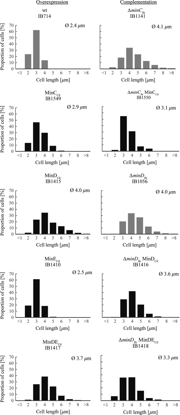
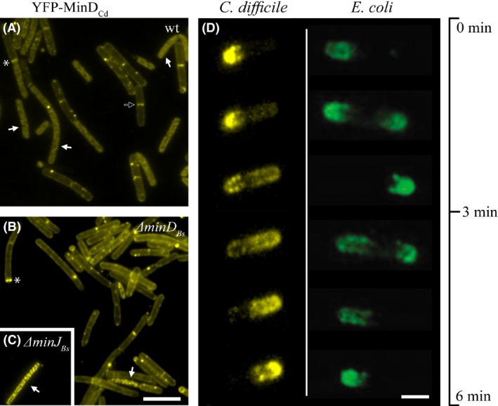
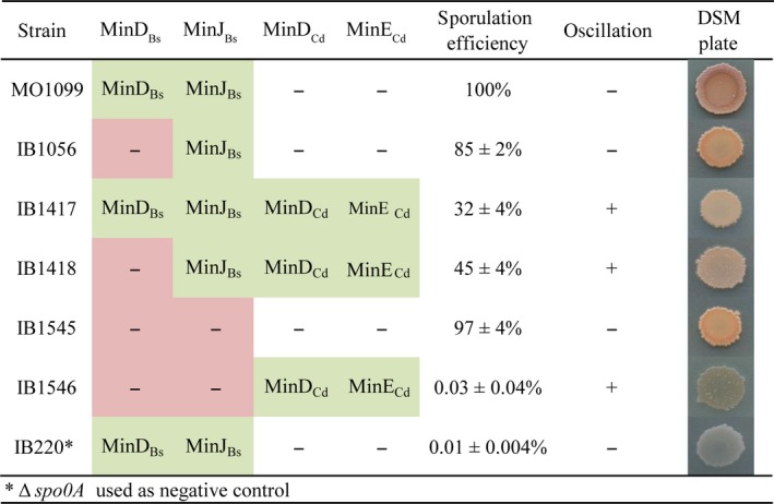
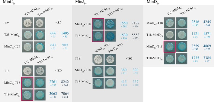
In rod-shaped bacteria, the proper placement of the division septum at the midcell relies, at least partially, on the proteins of the Min system as an inhibitor of cell division. The main principle of Min system function involves the formation of an inhibitor gradient along the cell axis; however, the establishment of this gradient differs between two well-studied gram-negative and gram-positive bacteria. While in gram-negative Escherichia coli, the Min system undergoes pole-to-pole oscillation, in gram-positive Bacillus subtilis, proper spatial inhibition is achieved by the preferential attraction of the Min proteins to the cell poles. Nevertheless, when E.coli Min proteins are inserted into B.subtilis cells, they still oscillate, which negatively affects asymmetric septation during sporulation in this organism. Interestingly, homologs of both Min systems were found to be present in various combinations in the genomes of anaerobic and endospore-forming Clostridia, including the pathogenic Clostridium difficile. Here, we have investigated the localization and behavior of C.difficile Min protein homologs and showed that MinDE proteins of C.difficile can oscillate when expressed together in B.subtilis cells. We have also investigated the effects of this oscillation on B.subtilis sporulation, and observed decreased sporulation efficiency in strains harboring the MinDE genes. Additionally, we have evaluated the effects of C.difficile Min protein expression on vegetative division in this heterologous host.

Keywords: Bacillus subtilis; Clostridium difficile; Min system oscillation; bacterial cell division; sporulation.


Figures

Figure 1
Figure 1
Cell length histograms. Left column: effects of overexpression of C. difficile Min proteins in wild‐type background. Expression was induced using 0.1 mM IPTG and/or 0.02% xylose, except for strain expressing MinCC d (IB1549), in which 0.3% xylose was used. Right column: complementation of MinBs proteins absence by MinCd proteins. Shown are induction conditions exhibiting the most notable complementation, that is 0.1 mM IPTG and/or 0.02% xylose except for strain ∆min DB s MinDEC d (IB1418), in which higher concentrations were used (0.5 mM IPTG and 0.3% xylose). Parental strains are in gray. Summary of all measurements can be found in Table S3.
Figure 2
Figure 2
Localization and oscillation of C.difficile Min proteins. (A)–(C) Localization of YFP‐tagged MinDC d expressed from Phyperspank in wild‐type and mutant B. subtilis backgrounds. (A) wt (IB1415), (B) ∆min DB s (IB1416), (C) ∆min JB s (IB1553). Full arrows point to examples of cells where the fine‐structure signal resembles the localization pattern of the native MinDB s along lipid spirals; the empty arrow indicates an example of localization to the vegetative septum, and the asterisk, to the asymmetric septum. Expression was induced with 0.1 mM IPTG; the scale bar represents 5 μm. (D) Time‐lapse images recorded over a period of 6 min showing oscillation of MinDC dMinEC d in a B.subtilis ∆min DB s background (IB1418), compared to oscillation of E.coli proteins in a B.subtilis ∆min DB s background (Jamroškovič et al. 2012). Expression of MinDC d was induced with 0.1 mM IPTG and MinEC d with 0.02% xylose; scale bar represents 1 μm. Available also as Video S1 in Supporting Information.
Figure 3
Figure 3
Sporulation efficiency of B.subtilis strains. Sporulation efficiency is given as the mean ± SD of at least three independent assays, each normalized against a wild‐type control. Sporulating colonies develop brown color, while nonsporulating light are brown to translucent, as seen in the ∆spo0A negative control (IB220).
Figure 4
Figure 4
Protein–protein interactions between MinDC d and the B.subtilis Min proteins compared alongside interactions among the B.subtilis Min proteins as detected by bacterial two‐hybrid system BACTH. Interactions were quantified using a β‐galactosidase assay and are expressed in Miller units as mean values ± SD of at least three independent measurements. The color intensity corresponds to the strength of the interaction; red boxes highlight strong positive interactions between heterologous proteins. Negative controls were all below 80 MU.
Figure 5
Figure 5
Bioinformatic analysis of MinCd proteins. (A) Percent sequence identity (same residues)/similarity (same residues + positive substitutions) between the Min proteins of B.subtilis (Bs), C.difficile (Cd) and E.coli (Ec) based on data from BLAST search (Altschul et al. 1997). (B) Model of the attachment of a MinDE c dimer to the membrane interface through its amphipathic helix. This model is based on the MinDE c crystal structure (PDB ID: 3Q9L), which contains residues 1–260, and thus lacks some of the residues involved in helix formation. Below: Alignment of the C‐terminal region of MinD containing the amphipathic helix. The consensus region is boxed, identical residues are violet, and conservation is indicated by color intensity.

Similar articles

Cited by

References

    1. Adams, D. W. , and Errington J.. 2009. Bacterial cell division, assembly, maintenance and disassembly of the Z ring. Nat. Rev. Microbiol. 7:642–653. - PubMed
    1. Adler, H. I. , Fisher W. D., Cohen A., and Hardigree A. A.. 1967. Miniature Escherichia coli cells deficient in DNA. Proc. Natl Acad. Sci. USA 57:321–326. - PMC - PubMed
    1. Altschul, S. F. , Madden T. L., Schäffer A. A., Zhang J., Zhang Z., Miller W., et al. 1997. Gapped BLAST and PSI‐BLAST: a new generation of protein database search programs. Nucleic Acids Res. 25:3389–3402. - PMC - PubMed
    1. Ausubel, F. M. , Brent R., Kingston R. E., Moore D. O., Seidmann J. S., Smith J., et al. 1987. Current Protocols in Molecular Biology. Wiley, New York.
    1. Barák, I. , and Wilkinson A. J.. 2007. Division site recognition in Escherichia coli and Bacillus subtilis . FEMS Microbiol. Rev. 31:311–326. - PubMed

MeSH terms

LinkOut - more resources