Review
. 2015 Dec 17;15 Suppl 1(Suppl 1):156.
doi: 10.1186/s12886-015-0132-x.

Meibomian gland dysfunction: hyperkeratinization or atrophy?

Affiliations
Review

Meibomian gland dysfunction: hyperkeratinization or atrophy?

James V Jester et al. BMC Ophthalmol. .

Abstract

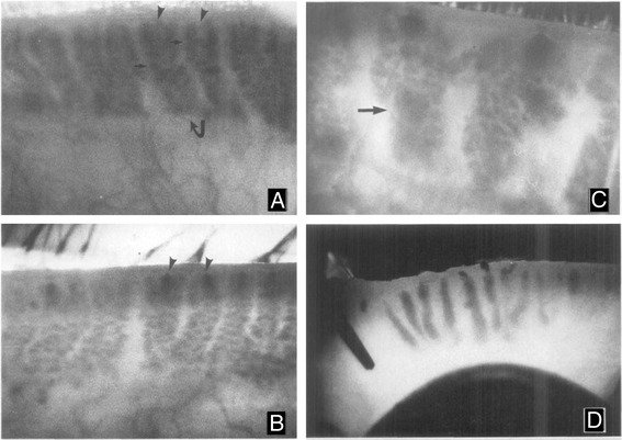
Meibomian gland dysfunction (MGD) is the major cause of evaporative dry eye disease (EDED) and dysfunction is widely thought to mechanistically involve ductal hyperkeratinization, plugging and obstruction. This review re-evaluates the role of hyperkeratinization in MGD based on more recent findings from mouse models. In these studies, eyelids from normal young and old mice or mice exposed to desiccating stress were evaluated by immunofluorescent tomography and 3-dimensional reconstruction to evaluate gland volume, expression of hyperkeratinization markers and cell proliferation or stimulated Raman scattering (SRS) microscopy to assess lipid quality. Results indicate that aging mice show dropout of meibomian glands with loss of gland volume and a forward migration of the mucocutaneous junction anterior to the gland orifice; similar age-related changes that are detected in human subjects. Atrophic glands also showed evidence of epithelial plugging of the orifice without the presence of hyperkeratinization. Mice exposed to desiccating stress showed hyperproliferation of the meibomian gland and ductal dilation suggesting a marked increase in lipid synthesis. Lipid quality was also affected in EDED mice with an increase in the protein content of lipid within the duct of the gland. Overall, age-related changes in the mouse show similar structural and functional correlates with that observed in clinical MGD without evidence of hyperkeratinization suggesting that gland atrophy may be a major cause of EDED. The response of the meibomian gland to desiccating stress also suggest that environmental conditions may accelerate or potentiate age-related changes.


Figures

Fig. 1
Fig. 1
Meibography of the rabbit eyelids from normal (a), and 2 % epinephrine treated eyes for 1–2 months (b), 3–4 months (c) and 6 months (d). Normal glands (a. curved arrow) appear as grape-like clusters of individual acini (small arrow) with indistinct gland orifices (a, arrowheads). After 1–2 months of treatment, the orifices of the glands become detectible as dark spots at the leading edge of the glands (b, arrowheads). Progression of hyperkeratinization into the gland is detected by 3–4 months after treatment as large, dark cystic structure that obscure the normal acini (c, arrows). After six months of treatment the meibomian glands are replaced by large, dense keratic cysts (d). Magnification A-C: 24x and D: 10x
Fig. 2
Fig. 2
Meibography of a patient suffering from chronic blepharitis (a) showing extensive meibomian gland acinar dropout compared to a normal individual (b)
Fig. 3
Fig. 3
Immunofluorescent Tomography of eyelids from 5 month-old (a, c, e) and 2 year-old (b, d, f) mice. In single plastic sections (a and b) sequentially stained for cytokeratin 1 (CK1, red), 5 (CK5, green) and 6 (CK6, blue) the mucocutaneous junction is detected by the transition from fully keratinized skin (CK1 positive) to non-keratinized conjunctiva (CK6 positive). Note the presence of ductal plugging with CK6 positive epithelial cells in the 2 year-old eyelid (arrowhead). Volume reconstruction of anti-CK5 staining of eyelids (c and d) with segmentation of the meibomian glands (green) from skin epithelium (white) shows marked atrophy of the meibomian glands in the 2 year-old compared to the 5 month-old eyelid (red arrowhead). Reconstruction of young and old eyelids (e and f) stained for CK1 epidermis (gold) and CK6 conjunctiva (green) show the anterior migration of the mucocutaneous junction (e and f, arrows)
Fig. 4
Fig. 4
Effects of desiccating stress on meibomian gland cell proliferation (a and b) and lipid quality (c-h). Cryosections stained for the cell cycling marker, Ki67 (green) of eyelids from normal mice (a) and mice exposed to 10 days of desiccating stress (b). SRS microscopy of lipid within cryosections from eyelids of normal mice (c) and the overlay (d) with immunostaining for nuclei (DAPI, red), and actin (Phalloidin, blue) to identify acini (ac, arrows) and ductules (dt). e, Graph of protein to lipid ratio from different regions of the gland (numbered 1–10), including acini (ac), ductule (dt) central duct (cd), and extracellular matrix (e). f-h, graph of protein to lipid ratio for control mice (f) and mice exposed to 5 days (g) and 10 days (h) desiccating stress (different colors represent different mice. Note that in the normal gland there is low glandular proliferation (a) and a gradual decreasing protein to lipid ratio moving from the acini to the central duct (e and f). By comparison meibomian glands from mice exposed to desiccating stress show up-regulation of cell cycling (b) and no change in the protein to lipid ratio (g and h)

Similar articles

Cited by

References

    1. Jester JV, Nicolaides N, Smith RE. Meibomian gland studies: histologic and ultrastructural investigations. Invest Ophthalmol Vis Sci. 1981;20:537–547. - PubMed
    1. Mishima S, Maurice DM. The oily layer of the tear film and evaporation from the corneal surface. Exp Eye Res. 1961;1:39–45. doi: 10.1016/S0014-4835(61)80006-7. - DOI - PubMed
    1. Hom MM, Martinson JR, Knapp LL, Paugh JR. Prevalence of Meibomian gland dysfunction. Optom Vis Sci. 1990;67:710–712. doi: 10.1097/00006324-199009000-00010. - DOI - PubMed
    1. Lemp MA, Nichols KK. Blepharitis in the United States, a survey-based perspective on prevalence and treatment. Ocul Surf. 2009;2009(7):S1–S14. doi: 10.1016/S1542-0124(12)70620-1. - DOI - PubMed
    1. Ong BL. Relation between contact lens wear and Meibomian gland dysfunction. Optom Vis Sci. 1996;73:208–210. doi: 10.1097/00006324-199603000-00015. - DOI - PubMed

Publication types