. 2016 Mar 18;291(12):6546-58.
doi: 10.1074/jbc.M115.705640. Epub 2016 Jan 27.

Ribosome Elongation Stall Directs Gene-specific Translation in the Integrated Stress Response

Affiliations

Ribosome Elongation Stall Directs Gene-specific Translation in the Integrated Stress Response

Sara K Young et al. J Biol Chem. .

Abstract

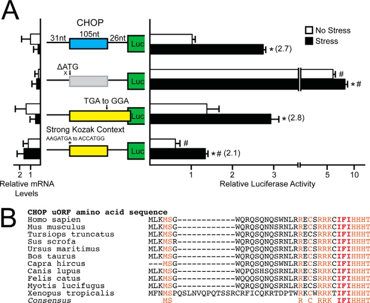
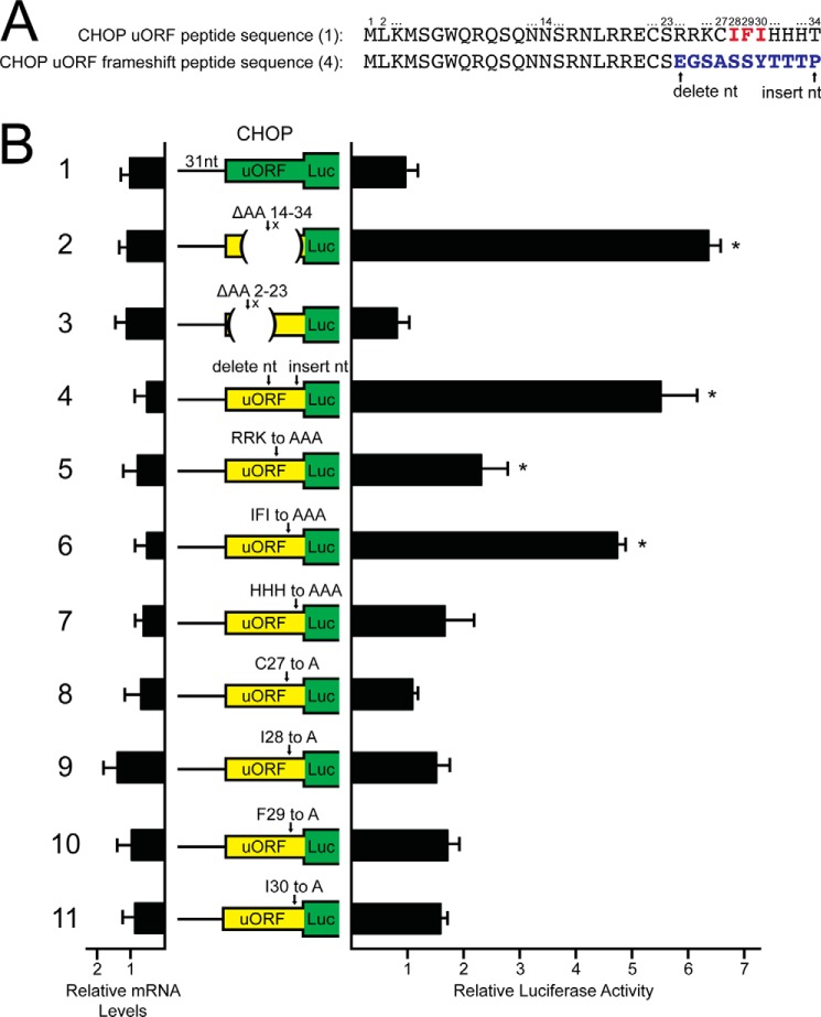
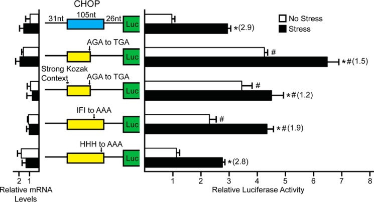
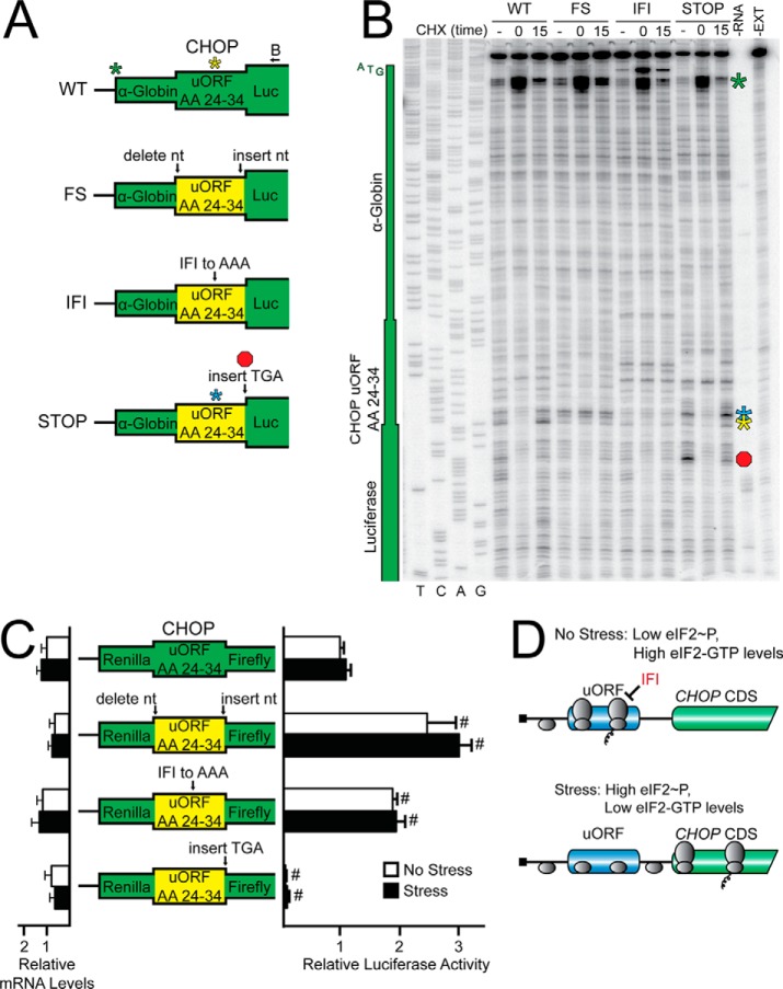
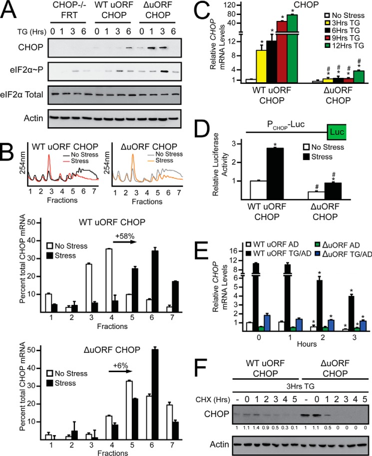
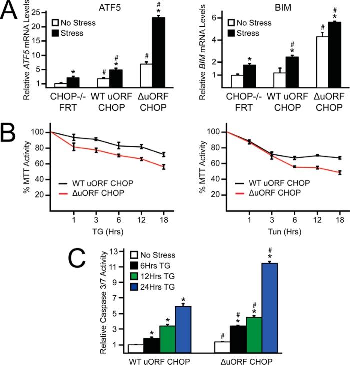
Upon exposure to environmental stress, phosphorylation of the α subunit of eIF2 (eIF2α-P) represses global protein synthesis, coincident with preferential translation of gene transcripts that mitigate stress damage or alternatively trigger apoptosis. Because there are multiple mammalian eIF2 kinases, each responding to different stress arrangements, this translational control scheme is referred to as the integrated stress response (ISR). Included among the preferentially translated mRNAs induced by eIF2α-P is that encoding the transcription factor CHOP (DDIT3/GADD153). Enhanced levels of CHOP promote cell death when ISR signaling is insufficient to restore cell homeostasis. Preferential translation of CHOP mRNA occurs by a mechanism involving ribosome bypass of an inhibitory upstream ORF (uORF) situated in the 5'-leader of the CHOP mRNA. In this study, we used biochemical and genetic approaches to define the inhibitory features of the CHOP uORF and the biological consequences of loss of the CHOP uORF on CHOP expression during stress. We discovered that specific sequences within the CHOP uORF serve to stall elongating ribosomes and prevent ribosome reinitiation at the downstream CHOP coding sequence. As a consequence, deletion of the CHOP uORF substantially increases the levels and modifies the pattern of induction of CHOP expression in the ISR. Enhanced CHOP expression leads to increased expression of key CHOP target genes, culminating in increased cell death in response to stress.

Keywords: CHOP; DDIT3; GADD153; endoplasmic reticulum stress (ER stress); eukaryotic initiation factor 2 (eIF2); stress response; translation control; translation initiation.


Figures

FIGURE 1.
FIGURE 1.
CHOP translation control involves bypass of an inhibitory uORF due in part to poor start codon context. A, WT and mutant versions of CHOP-Luc and a Renilla luciferase reporter were co-transfected into MEF cells and treated for 6 h with thapsigargin or left untreated. CHOP translation control was measured via a Dual-Luciferase assay and corresponding CHOP-Luc mRNA was measured by qRT-PCR. The CHOP-Luc construct contains the cDNA sequence corresponding to the CHOP 5′-leader fused to the luciferase reporter gene with the CHOP uORF and CDS of the CHOP-Luc fusion indicated with colored boxes. Mutant versions of CHOP-Luc include substitution of the CHOP uORF ATG start codons to AGG, mutation of the CHOP uORF stop codon from TGA to GGA, and optimization of the CHOP uORF start codons to the Kozak consensus sequence. Relative values are represented as histograms for each, with the S.D. indicated. The following values represent firefly luciferase activity normalized for mRNAs expressed from the indicated WT and mutant versions of CHOP-Luc reporters. These values feature the no stress values, stress values, and then in parentheses the induction ratios: WT, 1, 10.2 (10.2); ATG to AGG, 32.4, 24.1 (0.75); TGA to GGA, 0.9, 5.6 (6); and strong Kozak consensus, 0.5, 0.8 (1.6). B, polypeptide sequence encoded by the CHOP uORF from different vertebrates. The uORF polypeptide sequences were from cDNAs derived from CHOP orthologs, including Homo sapiens (BC003637), Mus musculus (BC013718), Tursiops truncates (XM_004316348), Sus scrofa (AK346731), Ursus maritimus (GW278660), Bos taurus (BC122721), Capra hircus (NM_001287231), Canis lupus (DN431044), Felis catus (XM_006933848), Myotis lucifugus (XM_006093575), and Xenopus tropicalis (BC153679). Residues conserved between the uORF sequences are listed in the consensus and are highlighted.
FIGURE 2.
FIGURE 2.
CHOP translation control involves an inhibitory uORF that relies on an encoded Ile-Phe-Ile sequence. A, representation of the CHOP uORF amino acid sequence in the wild-type context (construct 1) and the frameshift peptide sequence in which a nucleotide was deleted just after amino acid 23 and inserted following amino acid 34 (construct 4). The amino acid sequences in the uORF polypeptide are listed, with corresponding positions. The inhibitory Ile-Phe-Ile sequence is highlighted in red in the WT CHOP uORF peptide sequence. The last 10 amino acid residues in the CHOP uORF were altered as a consequence of the uORF frameshift construct and are highlighted in blue. B, the WT and mutant versions of the uORF-Luc constructs and a Renilla luciferase reporter were co-transfected into MEF cells. 24 h later, CHOP uORF translation control was measured via Dual-Luciferase assay, and the corresponding CHOP-Luc mRNAs were measured by qRT-PCR. The uORF-Luc constructs contain the CHOP uORF fused in-frame to the luciferase reporter gene, with the ATG start codon of luciferase deleted. The CHOP uORF sequence and luciferase CDS are indicated by the colored boxes. The green CHOP uORF box represents the wild-type CHOP uORF sequence. Yellow CHOP uORF boxes represent mutant constructs in which a change was made to the CHOP uORF sequence. Mutant versions of uORF-Luc include an in-frame deletion of uORF codons 14–34 or 2–23, frameshift in the last 10 CHOP uORF codons, substitution of CHOP uORF codons Arg-Arg-Lys to Ala-Ala-Ala, change of Ile-Phe-Ile to Ala-Ala-Ala, mutation of His-His-His to Ala-Ala-Ala, and alanine substitutions for Cys-27, Ile-28, Phe-29, and Ile-30. Relative values are represented as histograms for each, with the S.D. indicated. The following values represent firefly luciferase activity normalized for mRNA for the WT and mutant versions of the uORF-Luc reporters. The luciferase activity to mRNA ratios are: construct 1, 1; construct 2, 6.1; construct 3, 0.8; construct 4, 10.9; construct 5, 3; construct 6, 9.3; construct 7, 2.8; construct 8, 1.6; construct 9, 1.1; construct 10, 1.8; and construct 11, 1.9.
FIGURE 3.
FIGURE 3.
CHOP translation control involves bypass of an inhibitory uORF. WT and mutant versions of CHOP-Luc and a Renilla luciferase reporter were co-transfected into MEF cells and treated for 6 h with thapsigargin or left untreated. CHOP translation control was measured via a Dual-Luciferase assay, and corresponding CHOP-Luc mRNA was measured by qRT-PCR. The CHOP-Luc construct contains the cDNA sequence corresponding to the CHOP 5′-leader fused to the luciferase reporter gene with the CHOP uORF and CDS of the CHOP-Luc fusion indicated with colored boxes. Mutant versions of CHOP-Luc include mutation of the AGA codon of Arg-24 to TGA, simultaneous mutation of the CHOP uORF start codon to Kozak consensus sequence with the AGA to TGA mutation, change of the Ile-Phe-Ile codons to those encoding Ala-Ala-Ala, and mutation of the His-His-His codons to those encoding Ala-Ala-Ala. Relative values are represented as histograms for each, with the S.D. indicated. The following values represent firefly luciferase activity normalized for mRNA from the WT and mutant versions of CHOP-Luc reporters. The following features the no stress values, stress values, and then in parentheses the induction ratios: WT, 1, 1.8 (1.8); AGA to TGA, 2.5, 3.1 (1.2); optimized Kozak context with AGA to TGA, 3.8, 6.5 (1.7); IFI to AAA, 2.1, 4.1 (2); and HHH to AAA, 0.6, 2 (3.4).
FIGURE 4.
FIGURE 4.
Translation of the CHOP uORF results in a ribosome elongation stall that is dependent on an Ile-Phe-Ile sequence. A, depiction of toeprint design using the last 30 nucleotides of the CHOP uORF inserted in-frame between the rabbit α-globin and luciferase coding sequences to generate α-globin-CHOP-Luc fusion mRNA. Mutant versions of α-globin-CHOP-Luc mRNA include frameshift of the 30 nucleotides corresponding to the CHOP uORF (FS), mutation of the Ile-Phe-Ile codons to those encoding Ala-Ala-Ala (IFI), and insertion of a TGA stop codon just following the 30 CHOP uORF nucleotides (STOP). The black arrow depicted above the WT α-globin-CHOP-Luc mRNA represents the location of the primer used in B. Toeprints corresponding to ribosome initiation at the start codon for the WT and mutant α-globin-CHOP-Luc mRNAs are represented by a green star. Toeprints corresponding to an ribosome elongation stall for the WT, FS, and IFI mRNAs are represented by a yellow star. Toeprints corresponding to an elongation stall and ribosome termination for the STOP mRNA are represented by a blue star and a red octagon, respectively. B, cell-free translation extracts were treated with cycloheximide upon addition of WT or mutant versions of the α-globin-CHOP-Luc mRNA to measure initiating ribosomes (time 0), 15 min after addition of the transcript to measure ribosome localization during steady-state translation (time 15 min), or left untreated to measure prolonged ribosomal stalls that present without the use of an elongation inhibitor (time -). Toeprint assays were conducted for each sample, and sequencing reactions can be read 5′ to 3′ from top to bottom. The nucleotide complementary to the dideoxynucleotide added to each sequencing reaction is listed on the left, below the first four lanes. The products from control primer extension assays in the absence of RNA (−RNA) or in the absence of cell-free translation extracts are indicated on the right. The green star represents the toeprint corresponding to initiation at the α-globin-CHOP-Luc fusion, the yellow and blue stars represent prominent ribosome elongation stalls, and the red octagon represents the toeprint corresponding to termination at the introduced stop codon. The green boxes on the left span the sequences corresponding to the α-globin, CHOP uORF, and luciferase CDS and are comparable to the α-globin-CHOP-Luc schematic in A. Mutant constructs are the same listed in A, and the data are representative of three independent biological experiments. C, WT and mutant versions of Renilla-uORF-Luc were transfected into MEF cells and treated for 6 h with thapsigargin or left untreated. CHOP translation control was measured via a Dual-Luciferase assay and corresponding CHOP-Luc mRNA was measured by qRT-PCR. The Renilla-uORF-Luc construct includes the last 30 nucleotide residues of the CHOP uORF inserted in-frame between the Renilla and firefly luciferase coding sequences. Mutant versions of CHOP-Luc include frameshift of the 30 nucleotide segment corresponding to the CHOP uORF, mutation of the Ile-Phe-Ile codons to those encoding Ala-Ala-Ala, and insertion of a TGA stop codon just following the 30 nucleotide CHOP uORF segment. Relative values are represented as histograms for each, with the S.D. indicated. The following values represent firefly luciferase activity normalized for mRNA from the WT and mutant versions of CHOP-Luc reporters. The following features the no stress values, stress values, and then in parentheses the induction ratios: WT, 1, 0.9 (0.9); frameshift, 4, 4.1 (1); IFI to AAA, 1.6, 1.5 (0.9); and TGA insertion, 0.1, 0.1 (1). D, model for CHOP translation control. In the absence of stress, low eIF2α-P, and high eIF2-GTP, ribosomes scan the 5′-leader of the CHOP mRNA and initiate translation at the CHOP uORF. During translation of the uORF, elongating ribosomes are stalled at an Ile-Phe-Ile sequence, as depicted by the IFI sequence and black bar adjacent to the elongating ribosome in the uORF. The ribosome stall would preclude ribosome reinitiation downstream at the CHOP CDS. In the presence of stress and induced eIF2α-P, there would be lower levels of eIF2-GTP that would allow scanning ribosomes to bypass the CHOP uORF, in part because of its poor start codon context, and instead initiate translation at the CHOP CDS.
FIGURE 5.
FIGURE 5.
Alterations in CHOP uORF translation control change the dynamics of CHOP expression. A, MEF cells deleted for CHOP were stably selected to express WT CHOP (WT uORF CHOP) and CHOP with its uORF deleted (ΔuORF CHOP) and treated with the ER stress agent thapsigargin for up to 6 h or left untreated. The levels of CHOP, eIF2α-P, eIF2α total, and β-actin in these cultured cells were measured by immunoblot analyses. B, WT uORF CHOP and ΔuORF CHOP cells were treated with thapsigargin for 6 h or left untreated. Lysates were collected and layered on top of 10–50% sucrose gradients, followed by ultracentrifugation, and analysis of whole lysate polysome profiles at 254 nm. Sucrose gradients were fractionated simultaneous to analysis of polysome profiles at 254 nm. Total RNA was isolated from sucrose fractions and the percentage of total CHOP mRNA was determined by qRT-PCR. B is representative of three independent biological experiments. C, total RNA was collected from WT uORF CHOP and ΔuORF CHOP cells cultured in the presence or absence of thapsigargin and relative levels of CHOP mRNA were measured by qRT-PCR. D, fusion of 1 kb of the CHOP promoter (PCHOP-Luc) and a Renilla luciferase reporter were co-transfected into MEF cells, treated for 6 h or left untreated, and measured using a Dual-Luciferase assay. Relative values are represented as histograms, and the S.D. is indicated. E, total RNA was collected from WT uORF CHOP and ΔuORF CHOP cells cultured in the presence or absence of thapsigargin for 3 h followed by 0, 1, 2, or 3 h of treatment with actinomycin D. Relative levels of CHOP mRNA were measured by qRT-PCR. F, WT uORF CHOP and ΔuORF CHOP cells were treated with thapsigargin for 3 h, washed, and lysed (CHX −) or washed and treated with cycloheximide for up to 5 h (CHX 0, 1, 2, 3, 4, and 5). Levels of CHOP and β-actin in the cultured cells were measured by immunoblot analyses. Quantification of changes in CHOP protein expression are depicted under the CHOP immunoblot panel and are normalized to the no cycloheximide treatment for both WT uORF CHOP and ΔuORF CHOP cells.
FIGURE 6.
FIGURE 6.
Alterations in CHOP uORF translation control lower cell viability during stress. A, total RNA was collected from WT uORF CHOP and ΔuORF CHOP cells cultured in the presence or absence of thapsigargin. Relative levels of ATF5 and BIM mRNAs were measured in the cultured cells by qRT-PCR. B, equal numbers of WT uORF CHOP and ΔuORF CHOP cells were seeded in 96-well plates, cultured for 24 h, followed by treatment with thapsigargin or tunicamycin, as indicated, for up to an additional 18 h. MTT activity was measured by conversion of tetrazolium to formazan. C, equal numbers of WT uORF CHOP and ΔuORF CHOP cells were seeded in 96-well plates, cultured for 24 h, followed by treatment with or without thapsigargin for up to an additional 24 h. Caspase 3/7 activity was measured by cleavage of a proluminescent caspase-3/7 DEVD-aminoluciferin substrate.

Similar articles

Cited by

References

    1. Hinnebusch A. G. (2014) The scanning mechanism of eukaryotic translation initiation. Annu. Rev. Biochem. 83, 779–812 - PubMed
    1. Baird T. D., and Wek R. C. (2012) Eukaryotic initiation factor 2 phosphorylation and translational control in metabolism. Adv Nutr 3, 307–321 - PMC - PubMed
    1. Vattem K. M., and Wek R. C. (2004) Reinitiation involving upstream open reading frames regulates ATF4 mRNA translation in mammalian cells. Proc. Natl. Acad. Sci. U.S.A. 101, 11269–11274 - PMC - PubMed
    1. Palam L. R., Baird T. D., and Wek R. C. (2011) Phosphorylation of eIF2 facilitates ribosomal bypass of an inhibitory upstream ORF to enhance CHOP translation. J. Biol. Chem. 286, 10939–10949 - PMC - PubMed
    1. Young S. K., Willy J. A., Wu C., Sachs M. S., and Wek R. C. (2015) Ribosome reinitiation directs gene-specific translation and regulates the integrated stress response. J. Biol. Chem. 290, 28257–28271 - PMC - PubMed

Publication types

Substances